 Back
The Human Skeleton: Structure, Function, and Dynamics
Study Guide - Smart Notes
 Tailored notes based on your materials, expanded with key definitions, examples, and context.
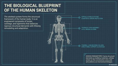
The Biological Blueprint of the Human Skeleton
Overview of the Skeletal System
The human skeleton provides the essential structural framework for the body, composed of bones, cartilage, and ligaments. This system is engineered to balance the demands of support, protection, and movement, while also adapting to lifelong remodeling and repair.
Cephalic Shield System: Offers complex cranial protection.
Thoracic Cage Assembly: Protects vital organs including the heart and lungs.
Femoral Load-Bearing Column: Provides structural support and acts as a lever for movement.
Design Parameters: Skeletal elements act as building materials, articulations as mechanical linkages, and cartilage as construction zones.
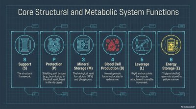
Core Structural and Metabolic System Functions
Six Primary Functions of the Skeleton
The skeletal system is multifunctional, serving both structural and metabolic roles essential for survival and homeostasis.
Support (S): Provides the main structural framework for the body.
Protection (P): Shields soft tissues and vital organs (e.g., brain, heart, lungs).
Mineral Storage (M): Acts as a reservoir for calcium and phosphorus, critical for metabolic processes.
Blood Cell Production (B): Hematopoiesis occurs in red marrow, producing red and white blood cells and platelets.
Leverage (L): Bones serve as rigid levers for muscle attachment, enabling movement.
Energy Storage (E): Yellow marrow stores triglycerides as an energy reserve.
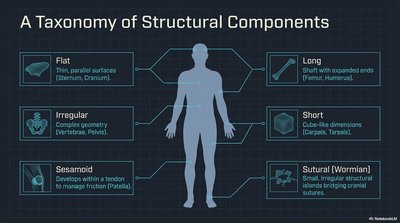
A Taxonomy of Structural Components
Classification of Bones by Shape
Bones are classified based on their shapes and structural roles, which reflect their functional adaptations.
Flat Bones: Thin, parallel surfaces (e.g., sternum, cranium).
Long Bones: Shaft with expanded ends (e.g., femur, humerus).
Short Bones: Cube-like dimensions (e.g., carpals, tarsals).
Irregular Bones: Complex geometry (e.g., vertebrae, pelvis).
Sesamoid Bones: Develop within tendons to manage friction (e.g., patella).
Sutural (Wormian) Bones: Small, irregular bones found in cranial sutures.
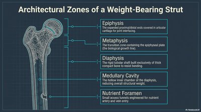
Architectural Zones of a Weight-Bearing Strut
Long Bone Structure
Long bones are organized into distinct anatomical zones, each with specialized functions for growth, support, and nutrient delivery.
Epiphysis: Expanded ends covered in articular cartilage for joint articulation.
Metaphysis: Transition zone containing the epiphyseal plate (growth region).
Diaphysis: The shaft, composed of thick compact bone for weight bearing.
Medullary Cavity: Hollow chamber within the diaphysis, housing marrow.
Nutrient Foramen: Small tunnels for nutrient artery and vein entry.
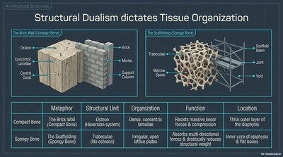
Structural Dualism Dictates Tissue Organization
Compact vs. Spongy Bone
Bones are composed of two main tissue types, each with distinct structural and functional properties.
Type | Metaphor | Structural Unit | Organization | Function | Location |
|---|---|---|---|---|---|
Compact Bone | The Brick Wall | Osteon (Haversian system) | Dense, concentric lamellae | Resists massive linear forces & compression | Thick outer layer of diaphysis |
Spongy Bone | The Scaffolding | Trabeculae | Irregular, open lattice | Absorbs multi-directional forces; structural weight reduction | Inner core of epiphyses & flat bones |
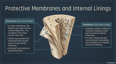
Protective Membranes and Internal Linings
Periosteum and Endosteum
Bones are covered and lined by specialized membranes that support growth, repair, and nutrient supply.
Periosteum (Outer Wrap): A two-layer membrane; the fibrous outer layer anchors tendons/ligaments, while the osteogenic inner layer houses stem cells. Highly vascularized and innervated, it is essential for bone repair.
Endosteum (Inner Lining): A delicate cellular membrane lining the medullary cavity and all internal trabeculae, containing osteoblasts and osteoclasts for ongoing remodeling.
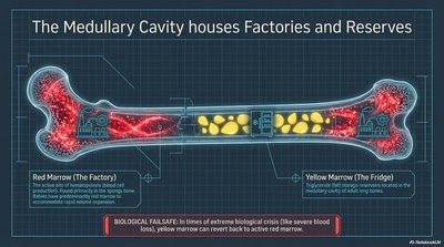
The Medullary Cavity: Factories and Reserves
Red and Yellow Marrow
The medullary cavity houses two types of marrow, each with distinct biological roles.
Red Marrow: Site of hematopoiesis (blood cell production), found in spongy bone of flat bones and epiphyses of long bones.
Yellow Marrow: Stores triglycerides (fat) and is found in the medullary cavity of adult long bones. In cases of severe blood loss, yellow marrow can revert to red marrow.
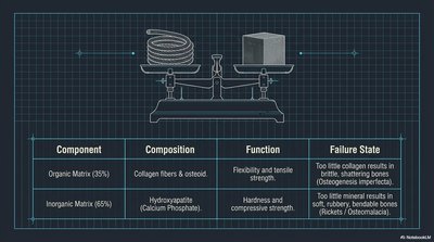
Bone Matrix Composition and Failure States
Organic and Inorganic Matrix
The bone matrix is a composite material, balancing flexibility and strength. Imbalances can lead to pathological conditions.
Component | Composition | Function | Failure State |
|---|---|---|---|
Organic Matrix (35%) | Collagen fibers & osteoid | Flexibility and tensile strength | Too little collagen: brittle, shattering bones (Osteogenesis imperfecta) |
Inorganic Matrix (65%) | Hydroxyapatite (Calcium Phosphate) | Hardness and compressive strength | Too little mineral: soft, bendable bones (Rickets/Osteomalacia) |
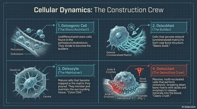
Cellular Dynamics: The Construction Crew
Bone Cell Types and Functions
Bone tissue is maintained and remodeled by four main cell types, each with specialized roles.
Osteogenic Cell (Stem/Architect): Undifferentiated stem cells found in periosteum and endosteum, giving rise to osteoblasts.
Osteoblast (Builder): Cells that secrete osteoid (unmineralized matrix) and build new bone tissue.
Osteocyte (Maintainer): Mature bone cells trapped in the matrix, maintaining bone tissue via cytoplasmic extensions in canaliculi.
Osteoclast (Demolition Crew): Large, multinucleated cells that resorb bone by secreting acids and enzymes, releasing calcium into the blood.
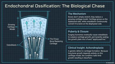
Endochondral Ossification: The Biological Chase
Bone Growth and Development
Most bones form through endochondral ossification, where cartilage is replaced by bone. This process is regulated by hormones and is essential for longitudinal growth.
Mechanism: Cartilage grows on the epiphyseal side, while osteoblasts replace it with bone on the diaphyseal side.
Puberty & Closure: Hormonal surges cause osteoblasts to outpace cartilage growth, closing the epiphyseal plate and ending longitudinal growth.
Clinical Insight: Achondroplasia results from a genetic defect in cartilage formation, leading to dwarfism due to early closure of growth plates.
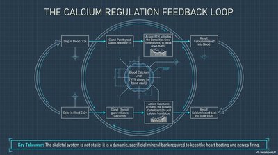
The Calcium Regulation Feedback Loop
Homeostasis of Blood Calcium
The skeletal system acts as a dynamic reservoir for calcium, regulated by hormonal feedback to maintain physiological balance.
Drop in Blood Ca2+: Parathyroid hormone (PTH) stimulates osteoclasts to release calcium from bone.
Spike in Blood Ca2+: Calcitonin from the thyroid inhibits osteoclasts and promotes calcium deposition in bone.
Key Takeaway: The skeleton is a dynamic mineral bank, essential for nerve and muscle function.
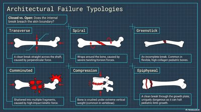
Architectural Failure Typologies
Types of Bone Fractures
Bones can fail in various ways depending on the force and direction of trauma. Understanding fracture types is crucial for clinical diagnosis and treatment.
Transverse: Clean break straight across the shaft.
Spiral: Caused by twisting forces, resulting in a spiral pattern.
Greenstick: Incomplete break, common in children.
Comminuted: Bone shatters into multiple fragments.
Compression: Bone is crushed under extreme vertical weight.
Epiphyseal: Break through the growth plate, dangerous for children.
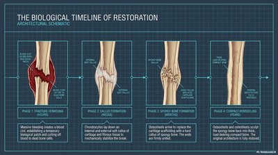
The Biological Timeline of Restoration
Bone Healing Process
Bone repair follows a predictable sequence of events, restoring structure and function after injury.
Phase 1: Fracture Hematoma – Blood clot forms, establishing a temporary scaffold for healing.
Phase 2: Callus Formation – Chondroblasts and osteoblasts form a soft callus of cartilage and fibrous tissue.
Phase 3: Spongy Bone Formation – Osteoblasts replace cartilage with spongy bone, stabilizing the fracture.
Phase 4: Compact Remodeling – Osteoclasts and osteoblasts remodel the bone, restoring original shape and strength.
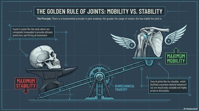
The Golden Rule of Joints: Mobility vs. Stability
Biomechanical Tradeoff
Joints are engineered to balance mobility and stability. The greater the range of motion, the less stable the joint, and vice versa.
Maximum Stability: Found in joints like the skull, which are immobile and protect vital organs.
Maximum Mobility: Found in joints like the shoulder, which allow extensive movement but are more prone to dislocation.
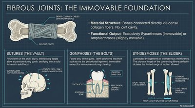
Fibrous Joints: The Immovable Foundation
Types and Functions
Fibrous joints are connected by dense collagen fibers and lack a joint cavity, resulting in little to no movement.
Sutures: Found only in the skull, interlocking edges provide stability.
Gomphoses: Peg-in-socket joints (e.g., teeth in alveolar sockets).
Syndesmoses: Bones connected by ligaments or interosseous membranes, allowing slight movement (e.g., between radius and ulna).
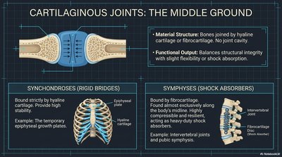
Cartilaginous Joints: The Middle Ground
Types and Functions
Cartilaginous joints unite bones with cartilage, providing both stability and limited flexibility.
Synchondroses: Bones joined by hyaline cartilage, typically immovable (e.g., epiphyseal plates).
Symphyses: Bones joined by fibrocartilage, allowing slight movement and shock absorption (e.g., intervertebral discs, pubic symphysis).
Synovial Joints: The Dynamic Apex
Structure and Function
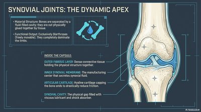
Synovial joints are the most mobile type, characterized by a fluid-filled cavity and specialized structures for movement and shock absorption.
Outer Fibrous Layer: Dense connective tissue holding the joint together.
Inner Synovial Membrane: Produces synovial fluid for lubrication.
Articular Cartilage: Hyaline cartilage capping bone ends to reduce friction.
Synovial Cavity: Space filled with viscous fluid for shock absorption.
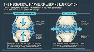
The Mechanical Marvel of Weeping Lubrication
Synovial Joint Lubrication Mechanism
Synovial joints utilize a unique lubrication system to withstand millions of movement cycles without cartilage degradation.
The Squeeze (Compression): Joint pressure forces synovial fluid out of cartilage, lubricating the surfaces.
The Soak (Relief): When pressure is relieved, cartilage reabsorbs fluid like a sponge, preparing for the next movement.
The Blueprint Synthesis: Where Form Meets Function
Joint Classification by Structure and Function
Joints are classified by both their structural material and their functional range of movement. This organization reflects evolutionary adaptations for protection, stability, and mobility.
Synarthroses (Immovable) | Amphiarthroses (Slightly Movable) | Diarthroses (Freely Movable) | |
|---|---|---|---|
Fibrous | Sutures (Skull), Gomphoses (Teeth) | Syndesmoses (Interosseous membranes) | |
Cartilaginous | Synchondroses (Costosternal) | Symphyses (Spine, Midline) | |
Synovial | All synovial joints (Knees, Shoulders, Limbs) |
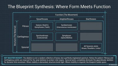
Key Master Insight: The skeleton is a perfectly engineered system, with fibrous and cartilaginous joints providing stability and protection, while synovial joints enable dynamic movement.
