 Back
The Immune System: Innate and Adaptive Defenses
Study Guide - Smart Notes
 Tailored notes based on your materials, expanded with key definitions, examples, and context.
The Immune System: Overview
The immune system is a complex network of cells, tissues, and molecules that defends the body against pathogens. It is divided into two main branches: innate immunity (nonspecific, immediate response) and adaptive immunity (specific, acquired response).
Innate Immunity
Barrier Defenses
Barrier defenses are the body's first line of defense, preventing pathogens from entering tissues.
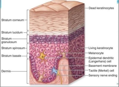
Skin: Acts as a physical barrier and produces secretions that inhibit or kill microbes (e.g., lysozyme in tears, low pH in sweat).
Mucous membranes: Trap pathogens and contain antimicrobial substances.
Secretions: Include enzymes and acidic environments that deter microbial growth.
Internal Defenses: Cellular Components
When pathogens breach barriers, internal cellular defenses are activated.
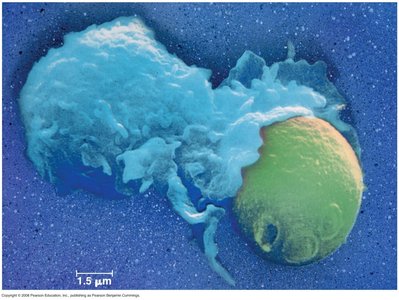
Phagocytes: White blood cells that engulf and digest pathogens.
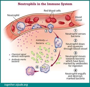
Neutrophils: Most abundant phagocytes (~60% of WBCs); rapidly respond to infection and enter tissues via diapedesis.
Monocytes: Largest WBCs (~6% of WBCs); differentiate into macrophages and dendritic cells.
Macrophages: Large phagocytes that reside in tissues and lymph nodes; can engulf many pathogens.
Dendritic cells: Specialized for antigen presentation; have long cytoplasmic extensions.
Recognition of Pathogens
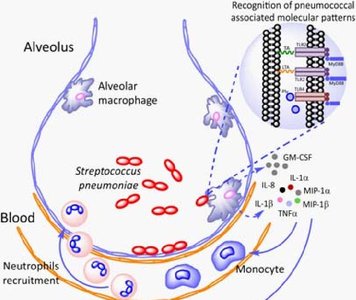
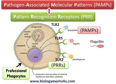
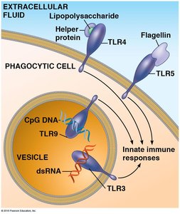
Innate immune cells recognize pathogens using pattern recognition receptors (PRRs) that bind to pathogen-associated molecular patterns (PAMPs) found on microbes.
Toll-like receptors (TLRs): A major class of PRRs in mammals; each TLR recognizes specific PAMPs (e.g., flagellin, lipopolysaccharide).
Binding of PAMPs to TLRs activates phagocytes and triggers the release of antimicrobial proteins.
The Lymphatic System
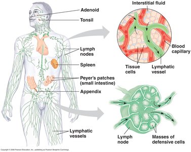
The lymphatic system supports immune function by transporting lymph and housing immune cells.
Lymph nodes: Sites where macrophages and lymphocytes interact with antigens.
Dendritic cells: Capture antigens in tissues and migrate to lymph nodes to activate adaptive immunity.
Other Cellular Defenses
Natural killer (NK) cells: Attack cells lacking class I MHC molecules, releasing perforins that lyse target cells.
Antimicrobial Proteins
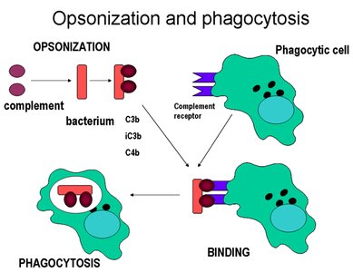
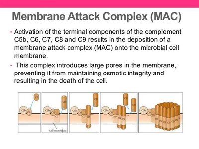
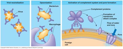
Complement system: Over 30 proteins that circulate in the blood in an inactive state. When activated, they enhance phagocytosis (opsonization) or directly lyse pathogens by forming the membrane attack complex (MAC).
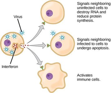
Interferons: Signaling proteins released by virus-infected cells; they induce resistance to viral replication in neighboring cells and activate immune cells.
Inflammation
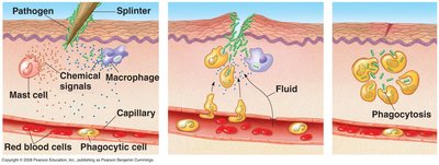
Inflammation is a localized response to infection or injury, characterized by heat, swelling, redness, and pain. Its main function is to recruit white blood cells to the site of infection.
Damaged cells release inflammatory chemicals.
Vasodilation and increased capillary permeability allow immune cells to access the affected tissue.
Phagocytes are attracted to the site and eliminate pathogens.
Adaptive (Acquired) Immunity
Adaptive immunity provides a specific response to pathogens and generates immunological memory. It involves lymphocytes (B cells and T cells) and antigen-presenting cells.
Key Terms
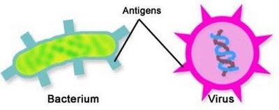
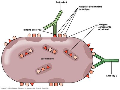
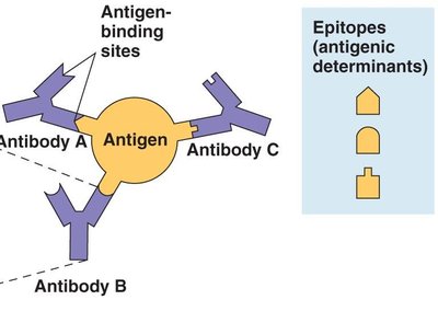
Antigen: A large molecule (often a protein or polysaccharide) recognized as "non-self" by the immune system; contains multiple epitopes (antigenic determinants).
Antibody: A protein produced by B cells that binds specifically to an antigen's epitope, marking it for destruction.
MHC proteins: Major histocompatibility complex molecules that present antigen fragments on cell surfaces.
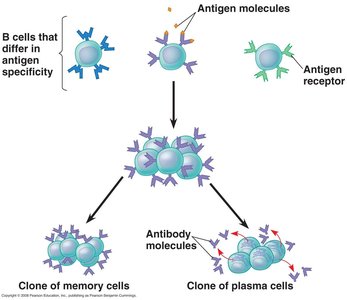
B cells: Lymphocytes that mature in bone marrow and produce antibodies (humoral immunity).
T cells: Lymphocytes that mature in the thymus; include helper T cells and cytotoxic T cells (cell-mediated immunity).
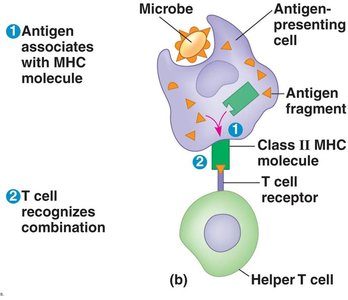
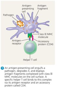
Antigen-presenting cells (APCs): Cells such as macrophages, dendritic cells, and B cells that display antigens via MHC II to activate T cells.
MHC Proteins
Class I MHC: Found on almost all nucleated cells; present endogenous antigens to cytotoxic T cells.
Class II MHC: Found mainly on APCs; present exogenous antigens to helper T cells.
Humoral Immune Response (B Cells)
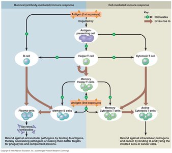
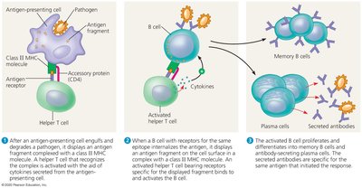
When a B cell encounters its specific antigen, it is activated (often with help from a helper T cell) and undergoes clonal selection, producing plasma cells (which secrete antibodies) and memory cells.
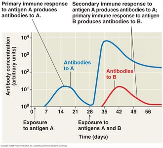
Primary response: First exposure to an antigen; antibody production peaks after about 10 days.
Secondary response: Subsequent exposures; faster and stronger antibody production due to memory cells.
Active and Passive Immunity
Active immunity: Body produces its own antibodies (via infection or vaccination).
Passive immunity: Antibodies are transferred from another source (e.g., maternal antibodies in breast milk, injection of antiserum).
Antibody Structure and Function
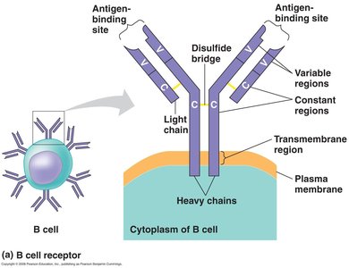
Antibodies (immunoglobulins) are Y-shaped proteins with variable regions for antigen binding and constant regions that determine class.
Antigen-antibody complexes can activate complement, neutralize pathogens, or enhance phagocytosis (opsonization).
Cell-Mediated Immunity (T Cells)
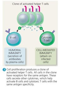
T cells are activated when their receptors bind to antigen-MHC complexes on APCs, with co-stimulation from helper T cells. Activated helper T cells secrete cytokines that stimulate B cells and cytotoxic T cells. Cytotoxic T cells destroy infected or abnormal cells displaying specific antigens.
Helper T cells: Activate B cells and cytotoxic T cells via cytokine secretion.
Cytotoxic T cells: Kill infected or cancerous cells presenting antigen with class I MHC.
Disruptions in Immune Function
Immunodeficiencies
Congenital immunodeficiency: Genetic defects impair immune cell function (e.g., SCID).
Acquired immunodeficiency: Caused by infections (e.g., HIV/AIDS) or treatments that suppress the immune system.
Autoimmune Diseases
The immune system attacks self-tissues, producing autoantibodies or self-reactive T cells.
Examples: Multiple sclerosis (myelin sheath destruction), rheumatoid arthritis (joint inflammation), type I diabetes (destruction of pancreatic islet cells).
Hypersensitivity
Immune response to harmless substances (e.g., pollen, animal dander) causes tissue damage (allergies).
Involves activation of mast cells and release of histamine upon re-exposure to allergen.
Additional info: This guide covers the essential mechanisms and components of the immune system, including both innate and adaptive branches, and highlights clinical implications such as immunodeficiencies, autoimmunity, and hypersensitivity reactions.
