Back
BackThe Integumentary System: Structure, Function, and Clinical Significance
Study Guide - Smart Notes
 Tailored notes based on your materials, expanded with key definitions, examples, and context.
The Integumentary System
Overview and Components
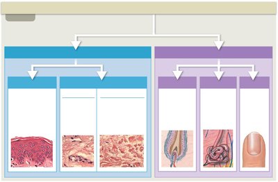
The integumentary system is the largest organ system in the human body, comprising approximately 16% of body weight and covering 1.5 to 2 m2 of surface area. It consists of two major parts: the cutaneous membrane (skin) and accessory structures such as hair, glands, and nails. The system also includes the subcutaneous layer (hypodermis), which lies beneath the dermis and consists of loose connective tissue.
Cutaneous membrane: Divided into the epidermis (outer layer) and dermis (inner layer).
Accessory structures: Originate in the dermis and extend through the epidermis to the skin surface (hair, exocrine glands, nails).
Subcutaneous layer: Provides insulation and anchors the skin to underlying tissues.
Functions of the Integumentary System
Protection: Shields underlying tissues and organs from environmental hazards.
Excretion: Removes salts, water, and organic wastes.
Thermoregulation: Maintains normal body temperature.
Melanin and Keratin Production: Provides UV protection and structural integrity.
Vitamin D3 Synthesis: Essential for calcium metabolism.
Lipid Storage: Energy reserve in the dermis and hypodermis.
Sensory Detection: Touch, pressure, pain, and temperature.
Immune Response Coordination: Defense against pathogens and cancers.
Epidermis
Structure and Cell Types
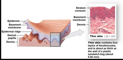
The epidermis is a stratified squamous epithelium that is avascular. Nutrients and oxygen diffuse from capillaries in the underlying dermis. The most abundant cell type is the keratinocyte, which produces keratin, a protein that provides strength and waterproofing.
Keratinocytes: Found on all exposed skin surfaces except the eyes.
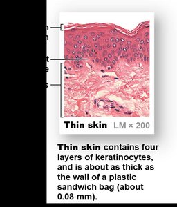
Thin skin: Covers most of the body; has four layers of keratinocytes.
Thick skin: Found on palms and soles; has five layers of keratinocytes.
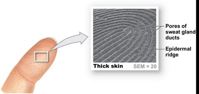
Strata of the Epidermis
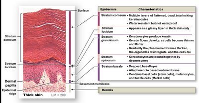
Thick skin contains five distinct layers (strata) of keratinocytes, arranged from the basement membrane to the free surface:
Stratum basale: Deepest layer; contains basal cells, melanocytes, and tactile cells.
Stratum spinosum: Keratinocytes are bound together by desmosomes.
Stratum granulosum: Keratinocytes produce keratin; cells become thinner and membranes thicken.
Stratum lucidum: Appears as a glassy layer in thick skin only.
Stratum corneum: Multiple layers of flattened, dead, interlocking keratinocytes; water resistant but not waterproof.
Dermis
Structure and Layers
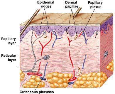
The dermis lies between the epidermis and the subcutaneous layer. It anchors epidermal accessory structures and consists of two main layers:
Papillary layer: Areolar tissue; contains capillaries, lymphatic vessels, and sensory neurons. Dermal papillae project between epidermal ridges.
Reticular layer: Dense irregular connective tissue; contains collagen and elastic fibers, and all cells of connective tissue proper.
Tension Lines (Cleavage Lines)
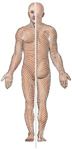
Tension lines are produced by parallel bundles of collagen and elastic fibers in the dermis. They resist forces applied to the skin. Surgical incisions made parallel to tension lines heal better and with less scarring than those made perpendicular.
Skin Color
Pigments and Blood Flow
Skin color is determined by two main pigments in the epidermis and by blood flow:
Melanin: Red-yellow or brown-black pigment produced by melanocytes; protects against UV radiation.
Carotene: Orange-yellow pigment from diet; accumulates in epidermal cells and can be converted to vitamin A.
Hemoglobin: Oxygenated blood gives skin a reddish hue; decreased oxygenation can cause cyanosis (bluish skin).
Accessory Structures
Hair and Hair Follicles
Hair and hair follicles are derived from the embryonic epidermis and located in the dermis. Hair protects the skull and provides sensory input.
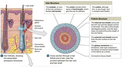
Hair structure: Medulla (core, soft keratin), cortex (thick layers, hard keratin), cuticle (outer layer, hard keratin).
Follicle structure: Internal and external root sheaths, glassy membrane, connective tissue sheath.
Exocrine Glands
The skin contains several types of exocrine glands:
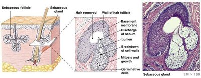
Sebaceous glands: Holocrine glands that secrete sebum (oil) into hair follicles; lubricates and protects hair shaft, inhibits bacterial growth.
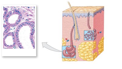
Apocrine sweat glands: Found in armpits, nipples, and pubic region; secrete sticky, odorous fluid into hair follicles.
Eccrine (merocrine) sweat glands: Widely distributed; discharge watery fluid directly onto skin surface; important for thermoregulation.
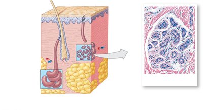
Mammary glands: Produce milk.
Ceruminous glands: Produce cerumen (earwax).
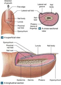
Nails
Nails protect the tips of fingers and toes and are composed of dead cells packed with keratin. Nail structure can be affected by metabolic disorders.
Nail body: Visible portion covering the nail bed.
Lateral nail grooves and folds: Surround the sides of the nail.
Hyponychium: Skin beneath the distal free edge.
Nail root: Site of nail production; visible nail emerges from under the eponychium (cuticle).
Lunula: Pale area near the root due to obscured blood vessels.
Repair and Aging of the Integument
Repair Mechanisms
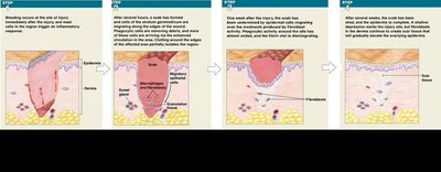
Following injury, the skin undergoes a series of repair processes:
Bleeding, swelling, and pain occur.
Mast cells trigger inflammation.
A scab forms to stabilize and protect the area.
Macrophages clean the area.
Fibroblasts and endothelial cells produce granulation tissue.
Effects of Aging
Epidermis thins.
Number of dendritic cells decreases.
Vitamin D3 production declines.
Melanocyte and glandular activity decreases.
Blood supply to dermis is reduced.
Hair follicle function declines.
Dermis thins and elastic fibers shrink.
Sex-specific hair and fat distribution fades.
Repair rate slows.
Integration with Other Body Systems
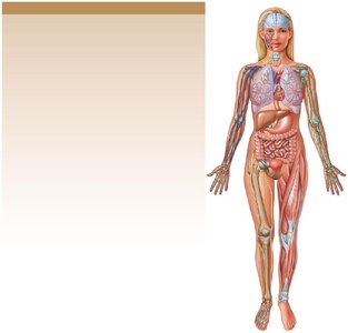
The integumentary system interacts with other body systems by providing protection, regulating temperature, synthesizing vitamin D3, storing lipids, detecting sensory stimuli, excreting wastes, and coordinating immune responses.