 Back
BackThe Mammalian Digestive System: Structure and Function
Study Guide - Smart Notes
 Tailored notes based on your materials, expanded with key definitions, examples, and context.
Organs Specialized for Sequential Stages of Food Processing in Mammals
Overview of the Mammalian Digestive System
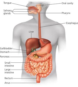
The mammalian digestive system is composed of the alimentary canal and various accessory glands that facilitate the breakdown and absorption of food. The process involves mechanical and chemical digestion, absorption of nutrients, and elimination of waste.
Alimentary canal: A continuous tube running from the mouth to the anus, including the oral cavity, pharynx, esophagus, stomach, small intestine, and large intestine.
Accessory glands: Salivary glands, pancreas, liver, and gallbladder secrete digestive juices that aid in food processing.
The Oral Cavity, Pharynx, and Esophagus
Initial Processing of Food
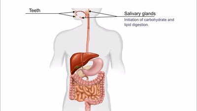
Food processing begins in the oral cavity, where mechanical and chemical digestion are initiated. Teeth cut and grind food, increasing its surface area for enzyme action. Saliva, secreted by salivary glands, contains mucus, buffers, antimicrobial agents, and the enzyme amylase, which begins the breakdown of starch and glycogen.
Mucus: Lubricates food, protects oral tissues, and aids in taste and smell.
Amylase: Initiates carbohydrate digestion.
Tongue: Shapes food into a bolus and assists in swallowing.
Swallowing and Passage to the Stomach
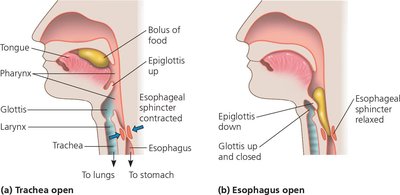
Swallowing moves the bolus from the oral cavity to the pharynx, a region that connects to both the esophagus and trachea. The epiglottis prevents food from entering the trachea during swallowing. Peristalsis, a series of muscle contractions, propels the bolus down the esophagus to the stomach.
Epiglottis: Flap that closes the trachea during swallowing to prevent choking.
Esophageal sphincter: Regulates entry of food into the stomach.
Digestion in the Stomach
Stomach Structure and Function
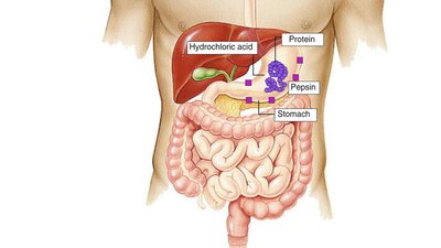
The stomach serves as a storage organ and a site for the mechanical and chemical breakdown of food. Its elastic walls allow it to hold up to 2 liters of food and fluid. Gastric juice, secreted by the stomach lining, contains hydrochloric acid (HCl) and the enzyme pepsin, which together convert food into a semi-liquid mixture called chyme.
HCl: Lowers pH to about 2, denatures proteins, and kills bacteria.
Pepsin: Protease that cleaves proteins into smaller polypeptides.
Churning: Muscular contractions mix food with gastric juice.
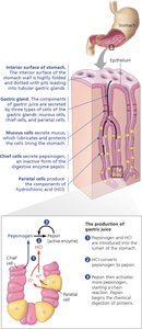
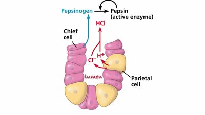
Gastric Glands and Positive Feedback
Gastric glands contain parietal cells (secrete HCl) and chief cells (secrete pepsinogen, the inactive precursor of pepsin). HCl activates pepsinogen to pepsin, and pepsin further activates more pepsinogen in a positive feedback loop. Mucus protects the stomach lining from self-digestion, and rapid cell turnover replaces damaged cells.
Equation for HCl formation:
Pepsinogen activation:
Digestion and Absorption in the Small Intestine
Chemical Digestion in the Small Intestine
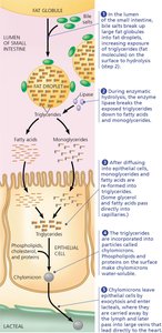
The small intestine is the primary site for enzymatic hydrolysis of macromolecules. The duodenum receives chyme from the stomach and digestive juices from the pancreas, liver, and gallbladder. Pancreatic enzymes (trypsin, chymotrypsin, amylase, lipase) and bile salts (from the liver, stored in the gallbladder) facilitate digestion of proteins, carbohydrates, nucleic acids, and fats.
Bicarbonate: Neutralizes acidic chyme.
Bile salts: Emulsify fats for efficient enzyme action.
Enzyme adaptation: Small intestine enzymes are adapted to neutral or slightly basic pH.
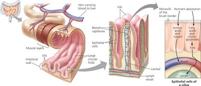
Absorption in the Small Intestine
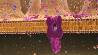
Most nutrient absorption occurs in the jejunum and ileum. The inner surface of the small intestine is highly folded and covered with villi and microvilli, greatly increasing surface area for absorption. Water-soluble nutrients (amino acids, sugars) enter the bloodstream, while fats are processed into chylomicrons and enter the lymphatic system.
Villi and microvilli: Increase surface area for absorption.
Hepatic portal vein: Transports nutrient-rich blood from the intestine to the liver for processing and detoxification.
Chylomicrons: Lipoprotein particles that transport fats via the lymphatic system.
Processing in the Large Intestine
Water Reabsorption and Feces Formation
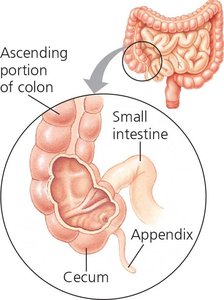
The large intestine (colon) absorbs water and forms feces from undigested material. The cecum, appendix, and colon are the main regions. The colon houses a large community of bacteria that aid in the breakdown of organic material and produce gases as metabolic by-products. The rectum stores feces until elimination through the anus.
Diarrhea: Results from insufficient water reabsorption.
Constipation: Results from excessive water reabsorption.
Appendix: Reservoir for symbiotic microorganisms.
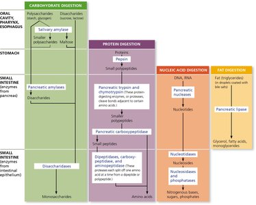
Summary Table: Digestive Enzymes and Their Actions
Location | Carbohydrate Digestion | Protein Digestion | Nucleic Acid Digestion | Fat Digestion |
|---|---|---|---|---|
Oral cavity, pharynx, esophagus | Salivary amylase | - | - | - |
Stomach | - | Pepsin | - | - |
Small intestine (from pancreas) | Pancreatic amylases | Trypsin, chymotrypsin, carboxypeptidase | Pancreatic nucleases | Pancreatic lipase |
Small intestine (from epithelium) | Disaccharidases | Dipeptidases, carboxypeptidase, aminopeptidase | Nucleotidases, nucleosidases, phosphatases | - |
Additional info: This table summarizes the main digestive enzymes, their sources, and the macromolecules they act upon throughout the digestive tract.
