 Back
Chapter 16 Biology
Study Guide - Smart Notes
 Tailored notes based on your materials, expanded with key definitions, examples, and context.
The Molecular Basis of Inheritance
Introduction
The molecular basis of inheritance centers on the role of DNA as the genetic material. This chapter explores the evidence supporting DNA's function, its structural features, the mechanisms of DNA replication, and the organization of chromosomes in both prokaryotic and eukaryotic cells.
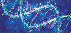
DNA as the Genetic Material
Historical Evidence for DNA's Role
Early experiments established DNA as the carrier of genetic information. T. H. Morgan's group showed genes are located on chromosomes, which are composed of DNA and protein. The role of DNA was further elucidated through studies of bacteria and viruses.
Griffith's Transformation Experiment
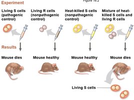
Frederick Griffith's 1928 experiment demonstrated that DNA can transform bacteria. He worked with two strains of Streptococcus pneumoniae: one pathogenic (S) and one harmless (R). Mixing heat-killed S cells with living R cells resulted in the transformation of R cells into pathogenic S cells, indicating a "transforming principle"—later identified as DNA.
Evidence from Bacteriophages
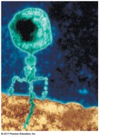
Bacteriophages (phages) are viruses that infect bacteria. Hershey and Chase's 1952 experiment used radioactive labeling to show that DNA, not protein, is the genetic material transmitted by phages to bacteria.
DNA Structure
Nucleotide Composition and Chargaff's Rules
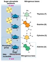
DNA is a polymer of nucleotides, each consisting of a nitrogenous base, a deoxyribose sugar, and a phosphate group. The four bases are adenine (A), thymine (T), guanine (G), and cytosine (C). Chargaff's rules state that the base composition of DNA varies between species, but the amount of A equals T and the amount of G equals C.
Base Pairing and Double Helix Structure
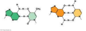
Watson and Crick determined that DNA is a double helix with specific base pairing: A pairs with T, and G pairs with C. This explains Chargaff's rules and provides a mechanism for DNA replication.
Hydrogen bonds stabilize base pairs: A-T pairs have two hydrogen bonds, G-C pairs have three.
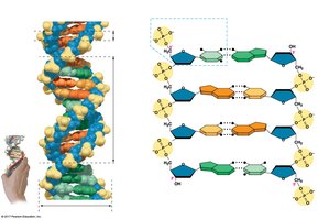
Antiparallel strands: DNA strands run in opposite directions (5' to 3' and 3' to 5').
X-ray Crystallography and DNA Structure
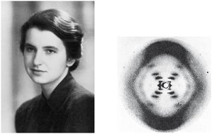
X-ray crystallography, used by Rosalind Franklin and Maurice Wilkins, provided critical evidence for the helical structure of DNA.
Double Helix Model
The double helix consists of two strands wound around each other, with bases paired in the interior and a sugar-phosphate backbone on the exterior. One full turn of the helix occurs every 10 base pairs.
DNA Replication
Mechanisms of DNA Replication
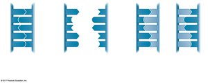
DNA replication is the process by which a cell copies its DNA before cell division. Each strand serves as a template for a new complementary strand. Three models were proposed: conservative, semiconservative, and dispersive. Meselson and Stahl's experiment confirmed the semiconservative model, where each new DNA molecule contains one old and one new strand.
Origins of Replication
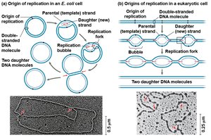
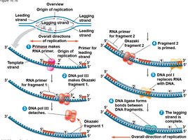
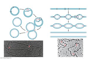
Replication begins at specific sites called origins of replication, forming replication bubbles that expand in both directions. In prokaryotes, there is typically one origin; in eukaryotes, there are multiple origins per chromosome.
Replication Fork and Enzymes
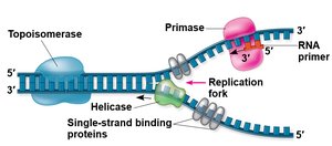
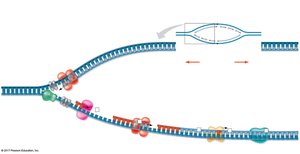
At the end of each replication bubble is a replication fork, where new DNA strands are synthesized. Key enzymes include:
Helicase: Unwinds the DNA helix.
Single-strand binding proteins: Stabilize unwound DNA.
Topoisomerase: Relieves strain ahead of the fork.
Primase: Synthesizes RNA primers.
Synthesizing New DNA Strands
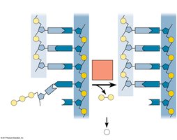
DNA polymerases catalyze the addition of nucleotides to the growing DNA strand. Each nucleotide added is a nucleoside triphosphate. DNA polymerases can only add nucleotides to the 3' end, so DNA elongates in the 5' to 3' direction.
Antiparallel Elongation: Leading and Lagging Strands
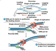
Due to the antiparallel nature of DNA, replication occurs differently on each strand:
Leading strand: Synthesized continuously toward the replication fork.
Lagging strand: Synthesized discontinuously away from the fork as Okazaki fragments, which are joined by DNA ligase.
Bacterial DNA Replication Proteins and Their Functions
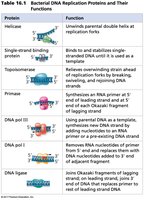
The main proteins involved in bacterial DNA replication and their functions are summarized below:
Protein | Function |
|---|---|
Helicase | Unwinds parental double helix at replication forks |
Single-strand binding protein | Stabilizes single-stranded DNA |
Topoisomerase | Relieves strain ahead of replication fork by breaking, swiveling, and rejoining DNA strands |
Primase | Synthesizes RNA primer at 5' ends of leading strand and Okazaki fragments |
DNA pol III | Using parental DNA as template, synthesizes new DNA strand by adding nucleotides to RNA primer or pre-existing DNA strand |
DNA pol I | Removes RNA nucleotides of primer and replaces them with DNA nucleotides |
DNA ligase | Joins Okazaki fragments of lagging strand; joins 3' end of DNA that replaces primer to rest of leading strand DNA |
Proofreading and Repairing DNA
DNA Polymerase Proofreading
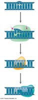
DNA polymerases proofread newly synthesized DNA, replacing incorrect nucleotides. Additional repair mechanisms include mismatch repair and nucleotide excision repair, which correct errors and remove damaged DNA segments.
Evolutionary Significance of Mutations
Although the error rate after proofreading and repair is low, mutations can occur. These sequence changes are the source of genetic variation, which drives evolution and the emergence of new species.
Chromosome Structure and DNA Packing
Bacterial Chromosome Organization
The bacterial chromosome is a double-stranded, circular DNA molecule associated with a small amount of protein. It is "supercoiled" and located in the nucleoid region of the cell.
Eukaryotic Chromosome Organization
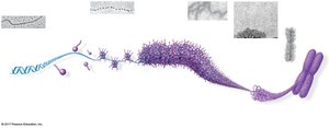
In eukaryotes, DNA is combined with proteins to form chromatin. Histones are responsible for the first level of DNA packing, forming nucleosomes. Chromatin is further organized into higher-order structures, ultimately forming condensed chromosomes during cell division.
Chromosome Territories in the Nucleus
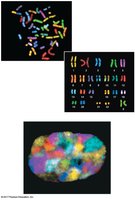
During interphase, chromosomes occupy specific regions within the nucleus, preventing entanglement and facilitating gene regulation.
Summary
DNA is the genetic material responsible for inheritance. Its double helix structure enables accurate replication, supported by a suite of enzymes. Chromosome organization varies between prokaryotes and eukaryotes, with complex packing mechanisms ensuring efficient storage and regulation of genetic information.
