 Back
Chapter 16 Study Guide
Study Guide - Smart Notes
 Tailored notes based on your materials, expanded with key definitions, examples, and context.
Chapter 16: The Molecular Basis of Inheritance
Introduction
This chapter explores the discovery, structure, and replication of DNA, the molecule responsible for storing and transmitting genetic information in living organisms. Understanding DNA's molecular basis is fundamental to genetics, molecular biology, and biotechnology.
Discovery of DNA as Genetic Material
Early Experiments Identifying Genetic Material
Griffith's Experiment (1928): Demonstrated that a "transforming principle" from dead pathogenic bacteria could make harmless bacteria pathogenic, suggesting the presence of an inheritable material.
Avery, McCarty, and MacLeod (1944): Identified DNA as the "transforming substance" responsible for heredity.
Hershey-Chase Experiment (1952)
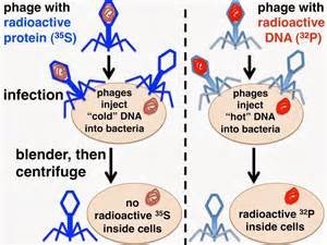
Hershey and Chase used bacteriophages (viruses that infect bacteria) to determine whether DNA or protein was the genetic material. They labeled viral DNA with radioactive phosphorus (32P) and viral protein with radioactive sulfur (35S). Only 32P was found inside infected bacteria, proving DNA is the genetic material.
Chargaff’s Rules
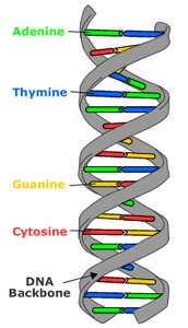
Erwin Chargaff (1950): Found that the amount of adenine (A) equals thymine (T), and guanine (G) equals cytosine (C) in DNA.
Base Pairing: A pairs with T, and G pairs with C, forming the basis for DNA's double helix structure.
Structure of DNA
Discovery of the Double Helix
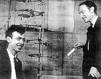
James Watson and Francis Crick (1953) built a model of DNA as a double helix, using data from Chargaff and X-ray diffraction images by Rosalind Franklin and Maurice Wilkins. Their model explained how DNA could replicate and carry genetic information.
Components of DNA
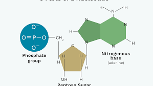
Nucleotide: The basic unit of DNA, consisting of a phosphate group, a deoxyribose sugar, and a nitrogenous base (A, T, G, or C).
Purines: Adenine (A) and Guanine (G) – double-ring structures.
Pyrimidines: Cytosine (C) and Thymine (T) – single-ring structures.
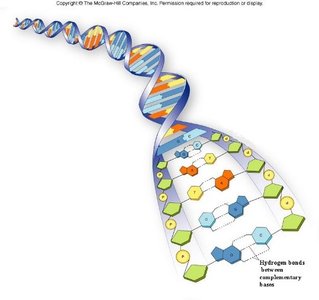
DNA Double Helix
Two strands run in opposite directions (antiparallel), forming a right-handed double helix.
The sugar-phosphate backbone is on the outside, with paired bases on the inside.
Hydrogen bonds hold the base pairs together: A-T (2 bonds), G-C (3 bonds).
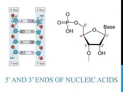
5’ and 3’ Ends of DNA
Each DNA strand has directionality, with a 5’ (phosphate) end and a 3’ (hydroxyl) end. DNA polymerases add nucleotides only to the 3’ end, determining the direction of synthesis.
DNA Replication
Overview of DNA Replication
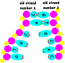
DNA replication is semiconservative: each new DNA molecule consists of one old (parental) strand and one newly synthesized strand.
Key Steps and Enzymes in DNA Replication
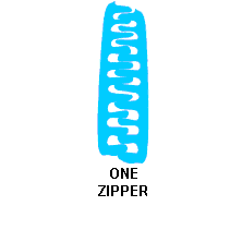
Helicase: Unwinds and separates the DNA strands.
Single-Strand Binding Proteins: Stabilize unwound DNA.
Topoisomerase: Relieves strain ahead of the replication fork by breaking, swiveling, and rejoining DNA strands.
Primase: Synthesizes a short RNA primer to provide a starting point for DNA synthesis.
DNA Polymerase: Adds nucleotides to the 3’ end of the primer, synthesizing the new DNA strand.
Complementary Base Pairing
New nucleotides are added according to base-pairing rules (A with T, G with C), ensuring accurate copying of genetic information.
Joining Complementary Bases
DNA polymerase catalyzes the formation of phosphodiester bonds between nucleotides, building the new DNA strand.
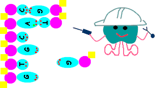
Semiconservative Replication
Each daughter DNA molecule contains one parental and one new strand, preserving genetic continuity.
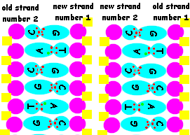
Antiparallel Elongation and Okazaki Fragments
Leading Strand: Synthesized continuously in the 5’ to 3’ direction.
Lagging Strand: Synthesized discontinuously as short Okazaki fragments, later joined by DNA ligase.
Proofreading and Repair of DNA
Proofreading: DNA polymerases correct errors during replication, resulting in high fidelity.
Mismatch Repair: Enzymes remove and replace incorrectly paired nucleotides.
Nuclease: Cuts out damaged DNA segments; DNA polymerase and ligase fill and seal the gap.
Humans have over 170 DNA repair enzymes, ensuring genome stability.
Telomeres
Telomeres are repetitive nucleotide sequences at the ends of eukaryotic chromosomes. They protect chromosome ends from deterioration and prevent activation of DNA damage responses. With each cell division, telomeres shorten, eventually leading to cell aging and division cessation.
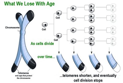
Chromosome Structure
Prokaryotes: DNA is circular and located in the cytoplasm.
Eukaryotes: DNA is linear and wraps around histone proteins to form nucleosomes, compacting into chromosomes.
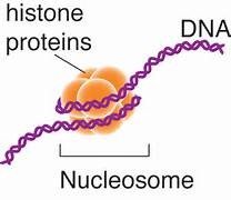
Summary Table: Key Experiments in DNA Discovery
Experiment | Year | Key Finding |
|---|---|---|
Griffith | 1928 | Transforming principle in bacteria |
Avery, McCarty, MacLeod | 1944 | DNA is the transforming substance |
Hershey-Chase | 1952 | DNA, not protein, is genetic material |
Chargaff | 1950 | Base pairing rules (A=T, G=C) |
Watson & Crick | 1953 | Double helix structure of DNA |
Key Equations
Base Pairing:
Directionality: DNA polymerase adds nucleotides to the 3' end:
