 Back
The Molecular Basis of Inheritance: Structure and Replication of DNA
Study Guide - Smart Notes
 Tailored notes based on your materials, expanded with key definitions, examples, and context.
The Molecular Basis of Inheritance
Introduction
The molecular basis of inheritance centers on the role of DNA as the genetic material. This chapter explores the experimental evidence for DNA as the hereditary molecule, its structure, and the mechanisms by which it is replicated and packaged within cells.
DNA as the Genetic Material
Historical Experiments Demonstrating DNA's Role
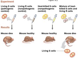
Griffith's Transformation Experiment (1928): Demonstrated that a substance from dead pathogenic bacteria could genetically transform living non-pathogenic bacteria, suggesting a "transforming principle."
Avery, MacLeod, and McCarty (1944): Identified DNA as the transforming principle.
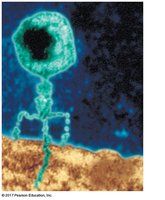
Hershey-Chase Experiment (1952): Used bacteriophages labeled with radioactive isotopes to show that DNA, not protein, is the genetic material injected into bacteria during viral infection.
Chargaff's Rules
Base Composition: The amount of adenine (A) equals thymine (T), and the amount of guanine (G) equals cytosine (C) in DNA.
Species Variation: The relative amounts of A, T, G, and C vary between species, supporting DNA's role in heredity.
Structure of DNA
DNA Nucleotide Structure
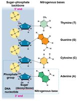
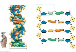
DNA is a polymer of nucleotides, each consisting of a nitrogenous base (A, T, G, or C), a deoxyribose sugar, and a phosphate group.
Double Helix Model
Watson and Crick (1953): Proposed the double helix structure of DNA, with two antiparallel strands held together by specific base pairing (A-T and G-C).
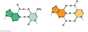
Base Pairing: Adenine pairs with thymine via two hydrogen bonds; guanine pairs with cytosine via three hydrogen bonds.
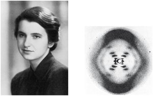
X-ray Crystallography: Rosalind Franklin's X-ray diffraction images were critical in elucidating the helical structure of DNA.
DNA Replication
Models of Replication
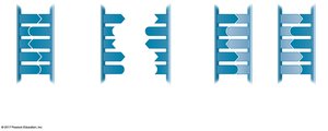
Semiconservative Model: Each new DNA molecule consists of one parental strand and one newly synthesized strand. Supported by Meselson and Stahl's experiment.
Other Models: Conservative and dispersive models were proposed but disproven.
Origins of Replication
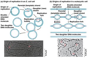
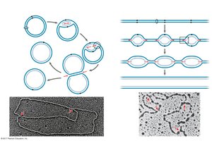
Replication begins at specific sites called origins of replication, forming replication bubbles that expand in both directions.
Enzymes and Proteins in DNA Replication
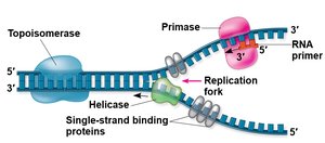
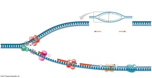
Helicase: Unwinds the DNA double helix at the replication fork.
Single-Strand Binding Proteins: Stabilize unwound DNA strands.
Topoisomerase: Relieves strain ahead of the replication fork by breaking and rejoining DNA strands.
Primase: Synthesizes short RNA primers needed to start DNA synthesis.
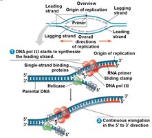
DNA Polymerase III: Adds nucleotides to the growing DNA strand in the 5' to 3' direction.
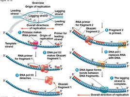
DNA Polymerase I: Removes RNA primers and replaces them with DNA.
DNA Ligase: Joins Okazaki fragments on the lagging strand.
Protein | Function |
|---|---|
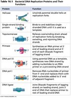
Helicase | Unwinds parental double helix at replication forks |
Single-strand binding protein | Binds to and stabilizes single-stranded DNA |
Topoisomerase | Relieves overwinding strain ahead of replication forks by breaking, swiveling, and rejoining DNA strands |
Primase | Synthesizes RNA primer at 5' end of leading strand and at 5' end of each Okazaki fragment of lagging strand |
DNA pol III | Using parental DNA as template, synthesizes new DNA strand by covalently adding nucleotides to the 3' end of a pre-existing DNA strand or RNA primer |
DNA pol I | Removes RNA nucleotides of primer from 5' end and replaces them with DNA nucleotides |
DNA ligase | Joins Okazaki fragments of lagging strand; on leading strand, joins 3' end of DNA that replaces primer to rest of leading strand DNA |
Leading and Lagging Strand Synthesis
Leading Strand: Synthesized continuously toward the replication fork.
Lagging Strand: Synthesized discontinuously away from the fork as Okazaki fragments, which are later joined by DNA ligase.
Proofreading and DNA Repair
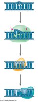
Proofreading: DNA polymerases correct errors during replication by removing mismatched nucleotides.
Mismatch Repair: Repair enzymes correct errors missed by DNA polymerase.
Nucleotide Excision Repair: Nucleases remove damaged DNA, which is then replaced by DNA polymerase and sealed by DNA ligase.
DNA Packaging in Chromosomes
Bacterial Chromosomes
Consist of double-stranded, circular DNA molecules associated with a small amount of protein.
DNA is supercoiled and located in the nucleoid region.
Eukaryotic Chromosomes and Chromatin Structure
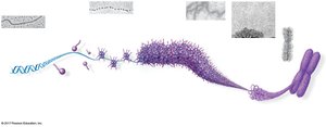
DNA is combined with proteins (histones) to form chromatin.
Chromatin undergoes multiple levels of packing: DNA double helix → nucleosomes → 30-nm fiber → looped domains → condensed chromosome.
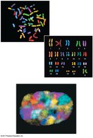
During interphase, chromosomes occupy specific regions in the nucleus and do not become entangled.
Summary Table: Key Concepts in DNA Structure and Replication
Concept | Key Points |
|---|---|
DNA as Genetic Material | Griffith, Avery, Hershey-Chase experiments; DNA, not protein, is hereditary material |
DNA Structure | Double helix, antiparallel strands, complementary base pairing (A-T, G-C) |
Replication | Semiconservative, origins of replication, leading/lagging strands, Okazaki fragments |
Enzymes | Helicase, primase, DNA polymerases, ligase, topoisomerase, single-strand binding proteins |
Repair | Proofreading, mismatch repair, nucleotide excision repair |
Chromosome Structure | Supercoiling in prokaryotes, chromatin packing in eukaryotes |
