 Back
The Molecular Basis of Inheritance: Structure, Replication, and Packaging of DNA
Study Guide - Smart Notes
 Tailored notes based on your materials, expanded with key definitions, examples, and context.
Chapter 16: The Molecular Basis of Inheritance
Introduction
This chapter explores the discovery, structure, replication, and packaging of DNA, the molecule responsible for the inheritance of genetic information in all living organisms. Understanding these processes is fundamental to modern biology and genetics.
DNA as the Genetic Material
The Search for the Genetic Material
Early 20th-century biologists debated whether DNA or protein was the genetic material. The role of DNA in heredity was first revealed through experiments with bacteria and viruses.
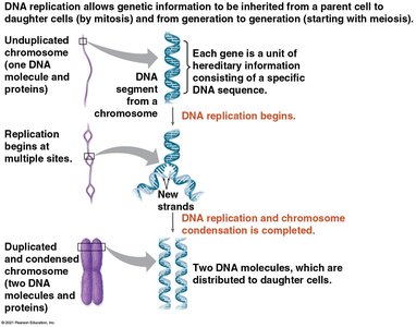
Genes are units of hereditary information located on chromosomes, which are composed of DNA and protein.
Key experiments with bacteria and viruses provided evidence that DNA, not protein, is the genetic material.
Evidence That DNA Can Transform Bacteria
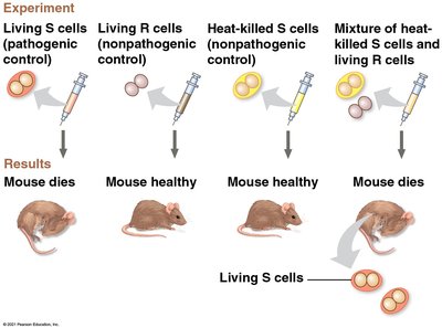
Frederick Griffith's 1928 experiment demonstrated that a substance from dead pathogenic bacteria could transform harmless bacteria into pathogenic forms, a process called transformation.
Transformation is now defined as a change in genotype and phenotype due to assimilation of foreign DNA.
Oswald Avery, Maclyn McCarty, and Colin MacLeod later identified DNA as the transforming substance.
Evidence That Viral DNA Can Program Cells
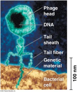
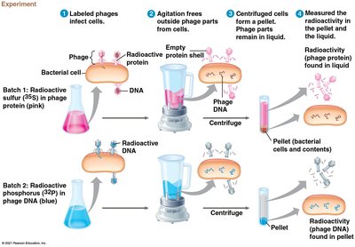
Bacteriophages (phages) are viruses that infect bacteria. Experiments by Hershey and Chase in 1952 showed that DNA, not protein, is the genetic material injected by phages into bacteria.
A virus consists of DNA (or RNA) enclosed in a protein coat.
Only DNA entered the bacterial cell and directed viral reproduction.
DNA Structure and Chargaff's Rules
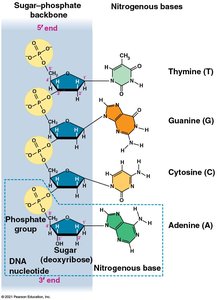
DNA is a polymer of nucleotides, each consisting of a nitrogenous base, a sugar, and a phosphate group. Erwin Chargaff discovered that DNA composition varies among species and that the amount of adenine equals thymine, and guanine equals cytosine (Chargaff's rules).
Nitrogenous bases: Adenine (A), Thymine (T), Guanine (G), Cytosine (C)
Chargaff's rules: A = T and G = C in any species
Discovery of the Double Helix
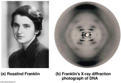
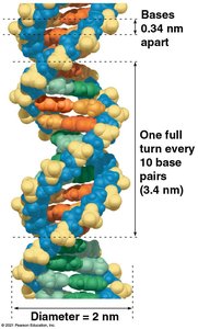
Maurice Wilkins and Rosalind Franklin used X-ray crystallography to study DNA structure. Franklin's images enabled Watson and Crick to deduce the double helical structure of DNA, with antiparallel sugar-phosphate backbones and specific base pairing.
DNA is composed of two antiparallel strands forming a double helix.
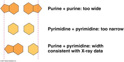
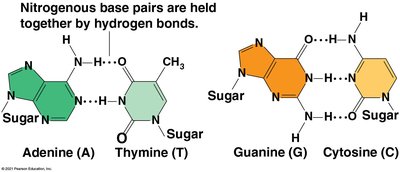
Base pairing: Purine (A or G) pairs with pyrimidine (T or C) for uniform width.
Hydrogen bonds hold base pairs together: A-T (2 bonds), G-C (3 bonds).
DNA Replication and Repair
Semiconservative Model of Replication
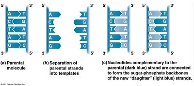
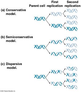
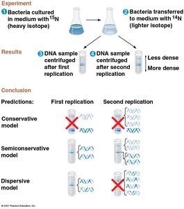
Watson and Crick proposed that DNA replication is semiconservative: each daughter molecule consists of one parental and one new strand. This was confirmed by Meselson and Stahl's experiments.
Alternative models: Conservative (parental strands rejoin) and dispersive (mix of old and new in each strand).
Mechanism of DNA Replication
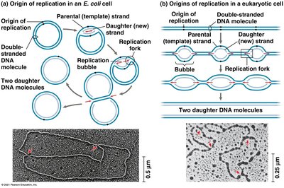
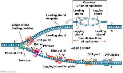
Replication begins at origins of replication, forming replication bubbles. Multiple enzymes and proteins coordinate the process.
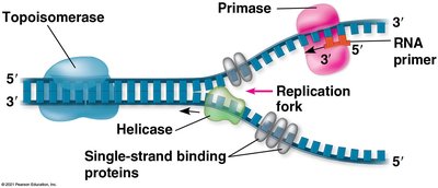
Replication fork: Y-shaped region where DNA is unwound.
Key enzymes: Helicase (unwinds DNA), single-strand binding proteins (stabilize unwound DNA), topoisomerase (relieves strain).
Synthesizing a New DNA Strand
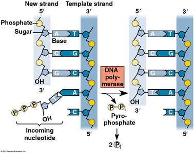
DNA polymerases synthesize new DNA by adding nucleotides to a primer. Synthesis occurs only in the 5' to 3' direction.
Primase synthesizes a short RNA primer.
DNA polymerase adds nucleotides to the 3' end of the primer.
Each nucleotide added is a nucleoside triphosphate (e.g., dATP).
Leading and Lagging Strands
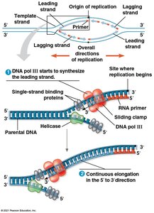
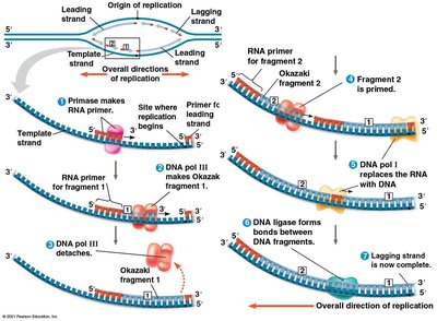
Due to the antiparallel nature of DNA, one strand (leading) is synthesized continuously, while the other (lagging) is synthesized in short fragments (Okazaki fragments) joined by DNA ligase.
Leading strand: Synthesized continuously toward the replication fork.
Lagging strand: Synthesized discontinuously away from the fork.
Okazaki fragments: Short DNA segments on the lagging strand.
Proteins Involved in DNA Replication
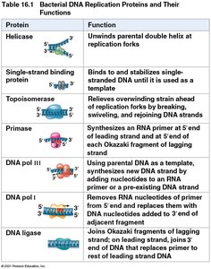
Multiple proteins coordinate the replication process, each with a specific function.
Protein | Function |
|---|---|
Helicase | Unwinds parental double helix at replication forks |
Single-strand binding protein | Binds to and stabilizes single-stranded DNA |
Topoisomerase | Relieves overwinding strain ahead of replication forks |
Primase | Synthesizes RNA primer at 5' end of leading strand and each Okazaki fragment |
DNA pol III | Uses parental DNA as template, synthesizes new DNA strand by adding nucleotides to RNA primer or pre-existing DNA strand |
DNA pol I | Removes RNA nucleotides of primer and replaces with DNA nucleotides |
DNA ligase | Joins Okazaki fragments and joins DNA that replaces primer to rest of leading strand DNA |
The DNA Replication Complex
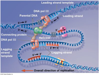
The proteins involved in DNA replication form a large complex, sometimes called the "DNA replication machine." DNA polymerases may remain stationary while DNA is threaded through the complex.
Proofreading and Repairing DNA
DNA polymerases proofread newly synthesized DNA, correcting errors. Additional repair mechanisms, such as mismatch repair and nucleotide excision repair, maintain DNA integrity.
Mismatch repair: Enzymes replace incorrectly paired nucleotides.
Nucleotide excision repair: Nucleases cut out and replace damaged DNA segments.
Mutations that escape repair can be inherited and are a source of genetic variation.
Replicating the Ends of DNA Molecules
Linear DNA in eukaryotes poses a challenge for replication at the ends (telomeres). Telomeres are repetitive sequences that protect genes from erosion. The enzyme telomerase extends telomeres in germ cells.
Telomere shortening is associated with aging and may protect against cancer by limiting cell divisions.
Telomerase activity is often detected in cancer cells, allowing them to divide indefinitely.
Chromosome Structure and DNA Packaging
DNA Packing in Chromosomes
Bacterial chromosomes are circular and supercoiled, while eukaryotic chromosomes are linear and highly organized with proteins. DNA and proteins form chromatin, which undergoes various levels of packing.
Histones: Main proteins responsible for DNA packing in eukaryotes.
Nucleosome: DNA wrapped twice around a core of eight histones.
Chromatin can be loosely packed (euchromatin) or highly condensed (heterochromatin).
Chromatin structure changes during the cell cycle, condensing into chromosomes for mitosis.
Regulation of Chromatin Structure
Histone modifications can alter chromatin condensation and affect gene expression. Chromatin organization is dynamic and essential for proper gene regulation and chromosome segregation.
