 Back
The Nervous System: Structure, Function, and Sensory Mechanisms
Study Guide - Smart Notes
 Tailored notes based on your materials, expanded with key definitions, examples, and context.
Nervous System Overview
Command and Control Center
The nervous system is responsible for gathering, processing, and organizing information, serving as the command and control center of the body. In humans, the brain contains approximately 100 billion neurons organized into complex circuits. Connections between brain regions can be mapped using the expression of colored proteins in neurons, revealing the intricate network of neural communication.
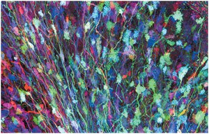
Neurons are specialized cells that transmit electrical and chemical signals.
Glial cells (glia) support, nourish, and regulate neurons.
The nervous system consists of circuits of neurons and supporting cells.
Evolution and Organization of Nervous Systems
Diversity of Nervous System Structures
Nervous systems vary widely among animal phyla, reflecting evolutionary adaptations to different lifestyles and body plans. Simple animals like cnidarians possess nerve nets, while more complex animals have centralized nervous systems with specialized structures.
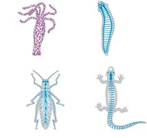
Nerve net: A diffuse network of interconnected nerve cells, as seen in cnidarians (e.g., Hydra).
Cephalization: Evolutionary trend toward clustering of sensory neurons and interneurons at the anterior end (head).
Central nervous system (CNS): Found in bilaterally symmetrical animals, consisting of a brain and longitudinal nerve cords.
Ganglia: Segmentally arranged clusters of neurons in annelids and arthropods.
Cells of the Nervous System
Neurons and Glia
Neurons are the primary signaling cells, while glial cells provide structural and metabolic support. In vertebrates, several types of glia are present, each with specialized functions.
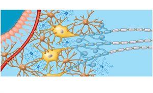
Oligodendrocytes (CNS) and Schwann cells (PNS): Form myelin sheaths around axons.
Astrocytes: Induce the formation of the blood-brain barrier and regulate the extracellular environment.
Microglia: Act as immune cells within the CNS.
Ependymal cells: Line the ventricles and help circulate cerebrospinal fluid.
Organization of the Vertebrate Nervous System
CNS and PNS Structure
The vertebrate nervous system is divided into the central nervous system (CNS) and the peripheral nervous system (PNS). The CNS includes the brain and spinal cord, while the PNS consists of nerves and ganglia outside the CNS.
Gray matter: Contains neuron cell bodies.
White matter: Composed of myelinated axons.
Cerebrospinal fluid: Circulates in the ventricles and central canal, providing nutrients and removing waste.
Peripheral Nervous System (PNS)
The PNS transmits information between the CNS and the rest of the body. It is divided into afferent (sensory) and efferent (motor) components.
Motor system: Controls voluntary and involuntary movements of skeletal muscles.
Autonomic nervous system: Regulates involuntary functions of smooth muscle, cardiac muscle, and glands. It has three divisions:
Sympathetic: "Fight-or-flight" responses.
Parasympathetic: "Rest-and-digest" functions.
Enteric: Controls digestive tract activity.
Development and Regionalization of the Brain
Embryonic Brain Regions
During embryonic development, the anterior neural tube differentiates into three primary brain regions: forebrain, midbrain, and hindbrain. These regions further subdivide and specialize in function.
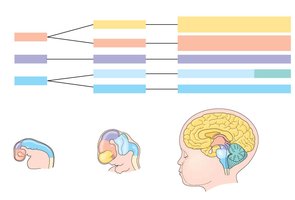
Forebrain: Gives rise to the cerebrum and diencephalon (thalamus, hypothalamus, epithalamus).
Midbrain: Part of the brainstem, involved in sensory processing.
Hindbrain: Forms the cerebellum, pons, and medulla oblongata.
Major Brain Structures and Functions
The vertebrate brain is regionally specialized, with each region responsible for distinct functions.
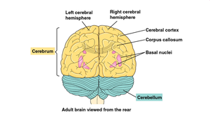
Cerebrum: Controls skeletal muscle contraction, learning, memory, and perception.
Cerebral cortex: Outer layer of the cerebrum, essential for higher cognitive functions.
Corpus callosum: Connects the two cerebral hemispheres.
Cerebellum: Coordinates movement and balance.
Brainstem and Diencephalon
The brainstem (midbrain, pons, medulla oblongata) is involved in basic life functions and information relay. The diencephalon includes the thalamus (sensory relay), hypothalamus (homeostasis, biological clock), and epithalamus.
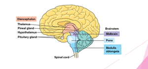
Medulla oblongata: Controls autonomic functions such as breathing and heart rate.
Pons: Relays information between the cerebrum and cerebellum.
Thalamus: Major sensory relay center.
Hypothalamus: Regulates temperature, hunger, thirst, and circadian rhythms.
Functional Systems of the Brain
Limbic System and Emotions
The limbic system, including the amygdala, hippocampus, and parts of the thalamus, is involved in the generation and experience of emotions. The amygdala is particularly important for emotional memory.
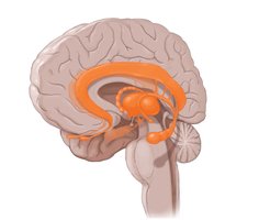
Limbic system: Integrates emotional responses with sensory input and memory.
Amygdala: Central to processing fear and emotional memories.
Cerebral Cortex and Cognitive Functions
The cerebral cortex is divided into four lobes (frontal, parietal, occipital, temporal), each associated with specific functions such as movement, sensation, language, and decision-making.
Frontal lobe: Decision making, planning, voluntary movement.
Parietal lobe: Sensory integration, touch perception.
Occipital lobe: Visual processing.
Temporal lobe: Auditory processing, language comprehension.
Lateralization of Brain Function
The left and right hemispheres of the cerebrum are specialized for different functions, a phenomenon known as lateralization. The corpus callosum enables communication between hemispheres.
Left hemisphere: Language, math, logic.
Right hemisphere: Spatial abilities, face recognition, nonverbal thinking.
Memory, Learning, and Neural Plasticity
Memory Formation
Memory involves changes in synaptic connections, a process known as neuronal plasticity. Short-term memory relies on temporary links in the hippocampus, while long-term memory involves stable connections in the cerebral cortex.
Hippocampus: Essential for forming new long-term memories.
Neuronal plasticity: The ability of the nervous system to change in response to experience.
Sensory and Motor Mechanisms
Sensory Reception and Transduction
Sensory receptors detect stimuli and convert them into electrical signals (receptor potentials). These signals are transmitted to the CNS, where they are interpreted as sensations.
Neuronal receptors: Sensory neuron acts as the receptor.
Non-neuronal receptors: Receptor cell releases neurotransmitter to a sensory neuron.
Receptor potential: Graded change in membrane potential in response to a stimulus.
Types of Sensory Receptors
Sensory receptors are classified by the type of stimulus they detect:
Mechanoreceptors: Detect pressure, touch, stretch, motion, and sound.
Electromagnetic receptors: Detect light, electricity, and magnetism.
Thermoreceptors: Detect temperature changes.
Pain receptors (nociceptors): Detect damaging or potentially damaging stimuli.
Chemoreceptors: Detect chemical stimuli (e.g., taste, smell).
Special Senses: Taste and Hearing
Taste (gustation) and smell (olfaction) rely on chemoreceptors. Hearing and equilibrium depend on mechanoreceptors that detect sound waves and body position.
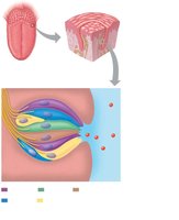
Taste buds: Contain sensory receptor cells for sweet, sour, salty, bitter, and umami.
Hearing: Involves the transduction of sound waves into nerve impulses by hair cells in the cochlea.
Equilibrium and Balance
Organs such as the utricle, saccule, and semicircular canals in the inner ear detect changes in head position and movement, helping maintain balance and spatial orientation.
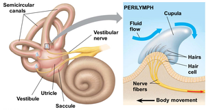
Otoliths: Granules in the utricle and saccule that detect gravity and linear acceleration.
Semicircular canals: Detect rotational movement.
Vision and Visual Processing
Structure of the Eye
The vertebrate eye is a single-lens organ that focuses light onto the retina, where photoreceptors (rods and cones) initiate visual transduction.
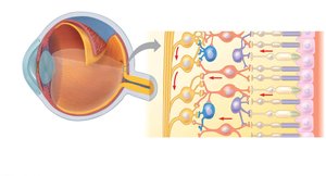
Cornea and lens: Focus light onto the retina.
Retina: Contains rods (sensitive to dim light) and cones (responsible for color vision).
Fovea: Region of highest visual acuity, densely packed with cones.
Phototransduction
Light absorption by photoreceptors triggers a cascade that converts cis-retinal to trans-retinal, activating rhodopsin and leading to changes in membrane potential and neurotransmitter release.
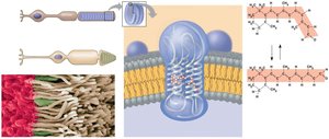
Rhodopsin: Visual pigment in rods, composed of opsin and retinal.
Photopsins: Visual pigments in cones, responsible for color vision.
Processing Visual Information
Visual information is processed in the retina and transmitted via the optic nerve to the brain, where it is interpreted in the visual cortex. The optic chiasm allows for the crossing of visual information from each eye to the opposite hemisphere.
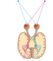
Receptive field: Area of the retina that influences the activity of a ganglion cell.
Optic chiasm: Site where optic nerve fibers cross, ensuring visual information from each field reaches the correct hemisphere.
Color Vision
Color vision in humans is based on three types of cones, each sensitive to different wavelengths (red, green, blue). Mutations in the genes encoding photopsins can lead to color blindness, which is more common in males due to X-linked inheritance.
Photopsins: Pigments in cones responsible for color discrimination.
Color blindness: Result of mutations in photopsin genes, often affecting red or green pigment.
Summary Table: Major Brain Regions and Functions
Region | Main Structures | Primary Functions |
|---|---|---|
Forebrain | Cerebrum, Diencephalon | Learning, memory, sensory processing, homeostasis |
Midbrain | Part of brainstem | Sensory integration, relay to forebrain |
Hindbrain | Cerebellum, Pons, Medulla | Movement coordination, autonomic functions |
Additional info: This guide integrates foundational concepts from Chapters 49 and 50, covering nervous system structure, function, sensory mechanisms, and the basics of sensory transduction and perception. For further study, refer to primary literature and advanced neuroscience texts.
