 Back
BackThe Respiratory System: Structure, Function, and Gas Exchange
Study Guide - Smart Notes
 Tailored notes based on your materials, expanded with key definitions, examples, and context.
The Respiratory System
Overview and Major Functions
The respiratory system is essential for supplying the body with oxygen (O2) required for cellular respiration and for removing carbon dioxide (CO2), a metabolic waste product. It also plays roles in olfaction (sense of smell) and speech production. The process of respiration involves both the respiratory and circulatory systems and can be divided into four main processes:
Pulmonary ventilation (breathing): Movement of air into and out of the lungs.
External respiration: Exchange of O2 and CO2 between the lungs and blood.
Transport: Movement of O2 and CO2 in the blood.
Internal respiration: Exchange of O2 and CO2 between systemic blood vessels and tissues.
Anatomy of the Respiratory System
Upper and Lower Respiratory Tracts
The respiratory system is divided into the upper respiratory tract and the lower respiratory tract:
Upper respiratory tract: Nose, nasal passages, sinuses, and pharynx.
Lower respiratory tract: Larynx, trachea, bronchi, bronchioles, lungs, and alveoli.
Upper Respiratory Tract: Structure and Function
The upper respiratory tract filters, warms, and humidifies incoming air. The nose and pharynx provide a passageway for respiration, house olfactory receptors, filter large particles and microorganisms, moisten and warm air, and serve as resonating chambers for the voice.
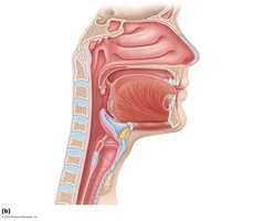
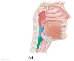
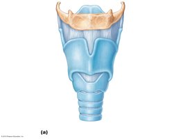
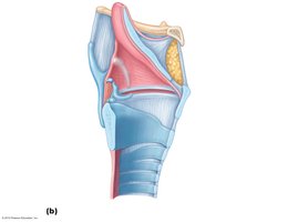
Lower Respiratory Tract: Structure and Function
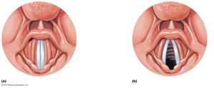
The lower respiratory tract is responsible for gas exchange and includes the larynx, trachea, bronchi, bronchioles, and alveoli. The larynx provides an open airway, routes air and food, and houses the vocal folds for sound production.
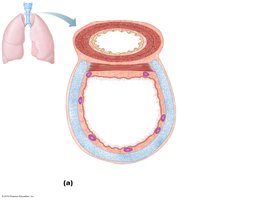
Trachea
The trachea (windpipe) transports air to and from the lungs. It is supported by C-shaped rings of cartilage and lined with ciliated, mucus-secreting epithelium that traps and removes foreign particles. The cough reflex helps expel irritants.
Bronchi and Bronchioles
The trachea branches into right and left bronchi, which further divide into smaller bronchioles. Bronchi contain cartilage and ciliated epithelium, while bronchioles lack cartilage. Both structures transport, clean, warm, and humidify air. Smoking damages cilia, leading to impaired clearance and smoker’s cough.
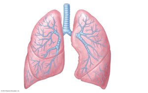
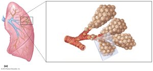
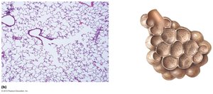
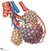
Lungs and Alveoli
The lungs occupy the thoracic cavity and are composed primarily of alveoli, the sites of gas exchange. The right lung has three lobes; the left lung has two lobes and a cardiac notch. The alveoli are tiny air sacs surrounded by capillaries, where O2 and CO2 are exchanged.
Mechanics of Breathing (Pulmonary Ventilation)
Inspiration and Expiration
Breathing consists of two phases:
Inspiration: Diaphragm and external intercostal muscles contract, increasing thoracic volume and decreasing intrapulmonary pressure, causing air to flow into the lungs.
Expiration: Muscles relax, thoracic volume decreases, intrapulmonary pressure rises, and air flows out of the lungs.
Pressure relationships are crucial: atmospheric pressure (Patm) is the reference point (760 mm Hg at sea level). Intrapulmonary and intrapleural pressures change during breathing, driving airflow.
Respiratory Volumes and Capacities
Respiratory volumes are used to assess lung function:
Tidal volume (TV): Normal breath (~500 mL).
Inspiratory reserve volume (IRV): Extra air inhaled after a normal inspiration.
Expiratory reserve volume (ERV): Extra air exhaled after a normal expiration.
Residual volume (RV): Air remaining after maximal exhalation.
Capacities are combinations of volumes (e.g., vital capacity, total lung capacity).
Dead Space
Anatomical dead space is the volume of air in the conducting passages (~150 mL) that does not participate in gas exchange. Alveolar dead space refers to non-functional alveoli. Total dead space is the sum of both.
Nonrespiratory Air Movements
Movements such as coughing, sneezing, laughing, and yawning modify normal respiratory rhythm and are often reflexive.
Gas Exchange and Transport
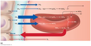
External and Internal Respiration
Gas exchange occurs by diffusion:
External respiration: O2 enters blood from alveoli; CO2 leaves blood for alveoli.
Internal respiration: O2 leaves blood for tissues; CO2 enters blood from tissues.
Oxygen Transport
O2 is transported in two forms:
1.5% dissolved in plasma
98.5% bound to hemoglobin (Hb) in red blood cells as oxyhemoglobin (HbO2)
Hemoglobin that has released O2 is called reduced hemoglobin (HHb).
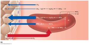
Carbon Dioxide Transport
CO2 is transported in three forms:
7–10% dissolved in plasma
20% bound to hemoglobin as carbaminohemoglobin
70% as bicarbonate ions (HCO3–) in plasma
The reaction for CO2 transport is:
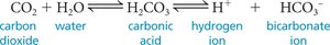
This reaction is catalyzed by the enzyme carbonic anhydrase in red blood cells.
Influence of CO2 on Blood pH
The carbonic acid–bicarbonate buffer system helps maintain blood pH. Changes in respiratory rate can adjust blood pH:
Slow, shallow breathing increases CO2, lowering pH (acidosis).
Rapid, deep breathing decreases CO2, raising pH (alkalosis).
Ventilation can compensate for metabolic disturbances in pH.
Respiratory Disorders Caused by Microorganisms
Common Respiratory Infections
Colds: Caused by over 100 viruses; symptoms include coughing, runny nose, congestion, and sneezing. Antibiotics are ineffective.
Influenza: Caused by influenza virus; symptoms include sore throat, fever, cough, aches, and chills. Pneumonia may develop, especially in vulnerable populations. Annual vaccination is recommended.
Pneumonia
Pneumonia is an infection of the lungs caused by bacteria or viruses. Alveoli fill with fluid, impairing gas exchange. Symptoms include fever, chills, shortness of breath, productive cough, and chest pain. Treatment depends on the cause.
Tuberculosis
Tuberculosis is a lung infection caused by Mycobacterium tuberculosis. It can be active or dormant. Diagnosis involves a skin test and chest X-ray; treatment requires antibiotics.
