 Back
The Structure and Function of Large Biological Molecules
Study Guide - Smart Notes
 Tailored notes based on your materials, expanded with key definitions, examples, and context.
The Molecules of Life
Overview of Biological Macromolecules
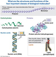
All living organisms are composed of four major classes of large biological molecules: carbohydrates, lipids, proteins, and nucleic acids. These molecules are essential for structure, function, and regulation of the body's cells, tissues, and organs.
Carbohydrates: Serve as fuel and building material.
Lipids: Hydrophobic molecules important for energy storage, membrane structure, and signaling.
Proteins: Perform a wide range of functions including catalysis, defense, transport, and structural support.
Nucleic acids: Store, transmit, and express hereditary information.
Macromolecules: Polymers and Monomers
Polymer Formation and Breakdown
Most biological macromolecules (except lipids) are polymers built from repeating units called monomers.
Polymer: A long molecule consisting of many similar or identical building blocks linked by covalent bonds.
Monomer: The repeating unit that serves as a building block for polymers.
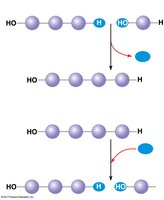
Dehydration reaction: Joins monomers by removing a water molecule, forming a new bond.
Hydrolysis: Breaks bonds between monomers by adding a water molecule.
Carbohydrates
Monosaccharides: Simple Sugars
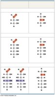
Carbohydrates include sugars and their polymers. The simplest carbohydrates are monosaccharides, or simple sugars, which generally have molecular formulas that are multiples of (CH2O)n.
Monosaccharides: Classified by the location of their carbonyl group (aldose or ketose) and the number of carbons in the skeleton (triose, pentose, hexose).
Glucose (C6H12O6): The most common monosaccharide.
Structure of Monosaccharides
Monosaccharides can exist in linear or ring forms. In aqueous solutions, many sugars form rings, which are energetically favorable. 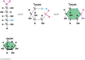
Disaccharides: Formation and Structure
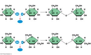
A disaccharide is formed when a dehydration reaction joins two monosaccharides. The covalent bond formed is called a glycosidic linkage.
Maltose: Formed from two glucose molecules (1–4 glycosidic linkage).
Sucrose: Formed from glucose and fructose (1–2 glycosidic linkage).
Polysaccharides: Storage and Structural Roles
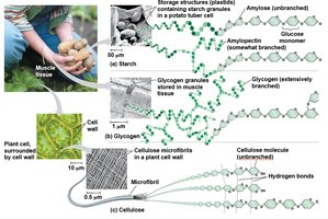
Polysaccharides are polymers of sugars and serve storage and structural functions.
Starch: Storage polysaccharide in plants, composed of glucose monomers.
Glycogen: Storage polysaccharide in animals, highly branched.
Cellulose: Structural polysaccharide in plant cell walls, composed of β glucose monomers.
Structural Differences: α and β Glucose
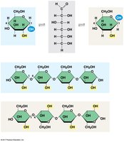
The structure of polysaccharides depends on the type of glucose monomer and the linkage.
Starch: 1–4 linkage of α glucose monomers.
Cellulose: 1–4 linkage of β glucose monomers.
Chitin: Structural Polysaccharide
Chitin is found in the exoskeleton of arthropods and the cell walls of many fungi. It is a structural polysaccharide similar to cellulose but contains nitrogen. 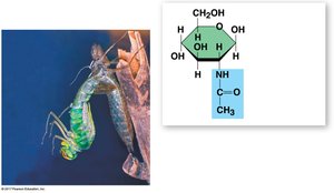
Lipids
Fats: Structure and Function
Lipids are hydrophobic molecules that do not form true polymers. The most important lipids are fats, phospholipids, and steroids.
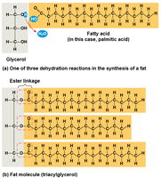
Fats: Constructed from glycerol and fatty acids. Major function is energy storage.
Glycerol: Three-carbon alcohol with a hydroxyl group attached to each carbon.
Fatty acid: Carboxyl group attached to a long hydrocarbon skeleton.
Saturated vs. Unsaturated Fats
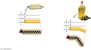
Saturated fats: No double bonds, solid at room temperature, mostly animal fats.
Unsaturated fats: One or more double bonds, liquid at room temperature, mostly plant and fish fats.
Phospholipids: Structure and Biological Role
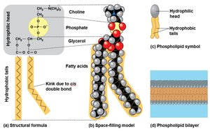
Phospholipids: Two fatty acids and a phosphate group attached to glycerol. Tails are hydrophobic; head is hydrophilic.
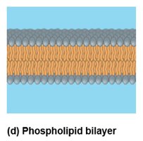
Form the phospholipid bilayer of cell membranes, creating a boundary between the cell and its environment.
Steroids: Structure and Function
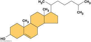
Steroids: Lipids with a carbon skeleton of four fused rings.
Cholesterol: Component of animal cell membranes and precursor for other steroids.
Proteins
Functions of Proteins
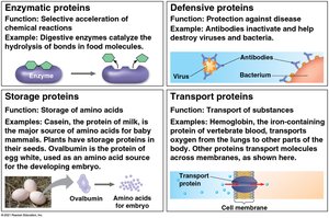
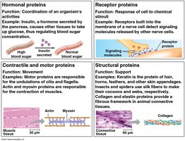
Proteins are the most diverse macromolecules, accounting for more than 50% of the dry mass of most cells. They perform a wide range of functions:
Enzymatic: Catalyze chemical reactions.
Defensive: Protect against disease.
Storage: Store amino acids.
Transport: Move substances.
Hormonal: Coordinate activities.
Receptor: Respond to chemical signals.
Contractile and motor: Movement.
Structural: Support.
Amino Acids: Building Blocks of Proteins
Proteins are constructed from 20 different amino acids. Each amino acid has an amino group, a carboxyl group, and a unique side chain (R group) that determines its properties. 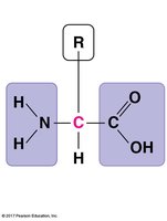
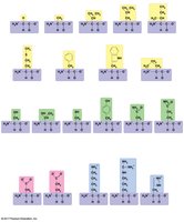
Polypeptides: Amino Acid Polymers
Amino acids are linked by peptide bonds to form polypeptides. Each polypeptide has a unique sequence, with an amino end (N-terminus) and a carboxyl end (C-terminus). 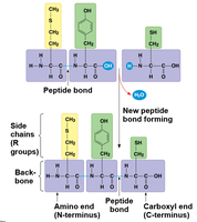
Protein Structure: Four Levels
The function of a protein depends on its three-dimensional structure, which is organized into four levels:
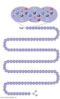
Primary structure: Unique sequence of amino acids.
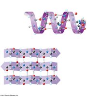
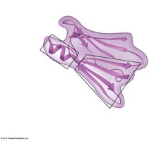
Secondary structure: Coils and folds (α helix, β pleated sheet) formed by hydrogen bonds.
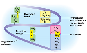
Tertiary structure: Overall shape determined by interactions among side chains (R groups).
Quaternary structure: Association of multiple polypeptide chains.
Protein Denaturation
Environmental changes such as pH, salt concentration, or temperature can cause proteins to lose their native structure, a process called denaturation. This results in loss of function. 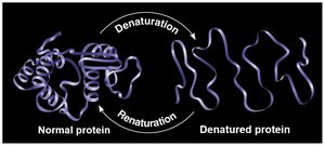
Nucleic Acids
Types and Functions
Nucleic acids are polymers called polynucleotides, made of monomers called nucleotides. There are two types:
DNA (Deoxyribonucleic acid): Stores genetic information and directs its own replication.
RNA (Ribonucleic acid): Functions in protein synthesis and gene expression.
Structure of Nucleic Acids
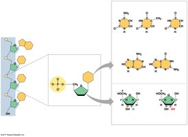
Nucleotide: Composed of a nitrogenous base, a pentose sugar (ribose or deoxyribose), and a phosphate group.
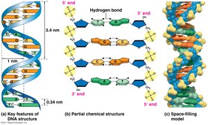
DNA: Double-stranded helix; two polynucleotides spiraling around an axis.
RNA: Single-stranded.
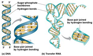
Key Features of DNA Structure
DNA strands are held together by hydrogen bonds between complementary bases (A-T, G-C).
DNA has a sugar-phosphate backbone and base pairs joined by hydrogen bonding.
 ----------------------------------------
