 Back
CH 5 PT 1 The Structure and Function of Large Biological Molecules: Carbohydrates and Proteins
Study Guide - Smart Notes
 Tailored notes based on your materials, expanded with key definitions, examples, and context.
The Molecules of Life
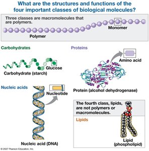
Overview of Biological Molecules
Biological molecules are essential for life, forming the structural and functional basis of cells. The four main classes are carbohydrates, proteins, nucleic acids, and lipids. Three of these classes are polymers, composed of repeating monomer units, while lipids are not true polymers.
Carbohydrates: Provide energy and structural support.
Proteins: Serve diverse functions including catalysis, structure, and transport.
Nucleic Acids: Store and transmit genetic information.
Lipids: Form membranes and store energy.
Foundations: Functional Groups
The Seven Functional Groups in Biological Molecules
Functional groups are specific groups of atoms within molecules that determine the chemical properties and reactivity of those molecules. They are found repeatedly in carbohydrates, proteins, nucleic acids, and lipids.
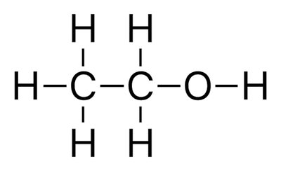
Hydroxyl (–OH): Polar, forms hydrogen bonds, found in sugars and alcohols.
Carbonyl (C=O): Polar, found in sugars (aldehyde or ketone forms).
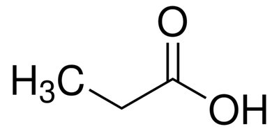
Carboxyl (–COOH): Acidic, donates H+, found in amino acids and fatty acids.
Amino (–NH2): Basic, accepts H+, found in amino acids.
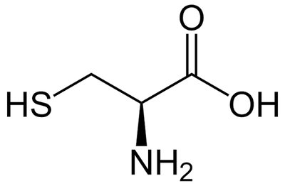
Sulfhydryl (–SH): Forms disulfide bonds, found in cysteine (protein structure).
Phosphate (–PO4): Acidic, negatively charged, found in nucleic acids and phospholipids.
Methyl (–CH3): Nonpolar, hydrophobic, found in fatty acids and gene regulation.
Monomers, Polymers, and Bonding
Polymer Formation and Breakdown
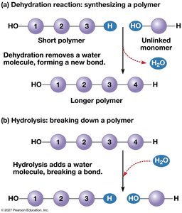
Biological macromolecules are formed by linking monomers into polymers via covalent bonds. The process of joining monomers is called dehydration synthesis, while breaking polymers into monomers is called hydrolysis.
Monomer: Small, repeating subunit (e.g., glucose, amino acid).
Polymer: Chain of monomers linked by covalent bonds.
Dehydration synthesis: Removes a water molecule to form a new bond.
Hydrolysis: Adds a water molecule to break a bond.
Carbohydrates
Monomers, Polymers, Bonds, and Elements
Carbohydrates are composed of carbon, hydrogen, and oxygen, typically in a 1:2:1 ratio. Their monomers are monosaccharides (simple sugars), which join to form polysaccharides via glycosidic bonds.
Monomer: Monosaccharide (e.g., glucose, fructose, galactose).
Polymer: Polysaccharide (e.g., starch, glycogen, cellulose, chitin).
Bond: Glycosidic bond (covalent bond linking sugar rings).
Elements: C, H, O (CH2O)n.


Functional Groups in Carbohydrates
Hydroxyl (–OH): Multiple per sugar; polar, water-soluble, forms glycosidic bonds.
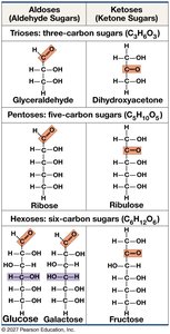
Carbonyl (C=O): One per sugar; defines sugar type (aldose or ketose).
Classification of Monosaccharides
Monosaccharides are classified by the number of carbons and the position of the carbonyl group.
Aldose: Carbonyl group at the end (aldehyde).
Ketose: Carbonyl group in the middle (ketone).
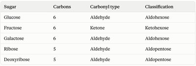
Sugar | Carbons | Carbonyl type | Classification |
|---|---|---|---|
Glucose | 6 | Aldehyde | Aldohexose |
Fructose | 6 | Ketone | Ketohexose |
Galactose | 6 | Aldehyde | Aldohexose |
Ribose | 5 | Aldehyde | Aldopentose |
Deoxyribose | 5 | Aldehyde | Aldopentose |
Formation of Disaccharides
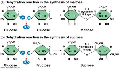
Disaccharides are formed by dehydration reactions between two monosaccharides, creating a glycosidic linkage.
Maltose: Glucose + Glucose (1-4 glycosidic linkage).
Sucrose: Glucose + Fructose (1-2 glycosidic linkage).
Comparison of Polysaccharides
Polysaccharides differ in their structure and function based on their monomer composition and branching.
Polysaccharide | Built from | Branching | Found in / Job |
|---|---|---|---|
Starch (amylose + amylopectin) | Glucose | Amylose: unbranched Amylopectin: branched | Plants — energy storage |
Glycogen | Glucose | Highly branched | Animals (liver/muscle) — energy storage |
Cellulose | Glucose (β-linkages) | Unbranched, linear chains | Plant cell walls — structural support |
Chitin | Modified glucose (+N group) | Unbranched, linear | Fungal cell walls & insect/arthropod exoskeletons — structural support |
Proteins
Amino Acids: Structure and Function
Proteins are polymers of amino acids, each with a central carbon bonded to an amino group, carboxyl group, hydrogen atom, and a variable R-group. The R-group determines the properties and function of each amino acid.
Amino group (–NH2): Basic, accepts protons.
Carboxyl group (–COOH): Acidic, donates protons.
R-group: Variable, determines chemical properties and folding.

R-Group Properties
Hydrophilic (polar): Attracted to water, often charged or contains O/N atoms.
Hydrophobic (non-polar): Repelled by water, mostly C and H, no charge.
Acidic R-group: Donates H+, becomes negatively charged.
Basic R-group: Accepts H+, becomes positively charged.
Levels of Protein Structure
Proteins fold into complex shapes, organized into four structural levels:
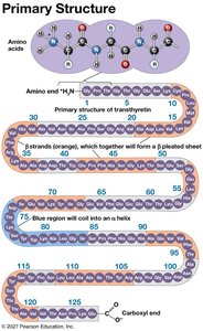
Primary structure: Linear sequence of amino acids, held by peptide bonds.
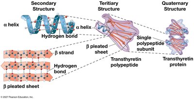
Secondary structure: Local folding into α-helices and β-sheets via hydrogen bonds.
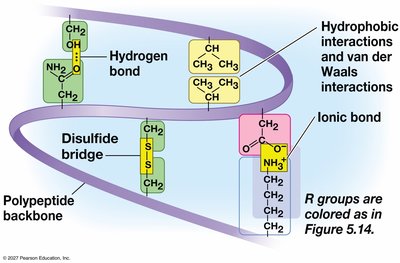
Tertiary structure: Overall 3D shape from R-group interactions (hydrophobic, ionic, hydrogen, disulfide bonds).
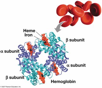
Quaternary structure: Assembly of multiple polypeptide subunits.
Bonds Responsible for Protein Structure
Level | Bond(s) Responsible |
|---|---|
Primary | Peptide bonds (covalent) between amino acids |
Secondary | Hydrogen bonds along the polypeptide backbone |
Tertiary | R-group interactions: hydrophobic clustering, ionic bonds, hydrogen bonds, disulfide bonds |
Quaternary | Same R-group interactions as tertiary, but between separate subunits |
Environmental Effects on Protein Structure
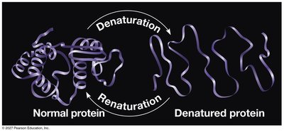
Protein structure and function are sensitive to environmental conditions such as temperature and pH. These factors disrupt weak bonds, altering protein shape and function.
Conformational change: Normal, reversible shift in shape; primary structure remains intact.
Denaturation: Abnormal, often irreversible loss of higher-level structure; protein unfolds and loses function, but peptide bonds are not broken.
Functions of Proteins in Cells
Proteins serve a wide range of functions in biological systems:
Enzymes: Catalyze chemical reactions.
Structural: Provide support and shape (e.g., collagen, keratin).
Transport: Move substances (e.g., hemoglobin carries O2).
Defense: Protect against pathogens (e.g., antibodies).
Movement: Enable contraction (e.g., actin, myosin).
Signaling: Communicate information (e.g., hormones like insulin).






Biological Structure and Function
The principle that structure determines function applies at all levels of biological organization, from protein structure to cells, tissues, organs, and organ systems.
Example: Actin/myosin protein structure enables muscle cell contraction, which leads to muscle tissue shortening and movement of the musculoskeletal system.
Elements Found in Proteins
Proteins are composed of carbon, hydrogen, oxygen, nitrogen, and sulfur. Sulfur is unique to proteins, found in the R-groups of cysteine and methionine, enabling disulfide bonds.
Carbon (C)
Hydrogen (H)
Oxygen (O)
Nitrogen (N)
Sulfur (S)





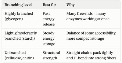
Summary Table: Branching in Polysaccharides
Branching Level | Best For | Why |
|---|---|---|
Highly branched (glycogen) | Fast energy release | Many free ends = many enzymes working at once |
Lightly/moderately branched (starch) | Steady energy storage | Balance of accessibility and compact storage |
Unbranched (cellulose, chitin) | Structural strength | Straight chains pack tightly and H-bond into strong fibers |
Additional info: Nucleic acids and lipids are briefly mentioned but not covered in detail in these notes. For a complete understanding, refer to textbook chapters on nucleic acids and lipids.
