 Back
BackThe Structure and Function of Large Biological Molecules: Proteins and Nucleic Acids
Study Guide - Smart Notes
 Tailored notes based on your materials, expanded with key definitions, examples, and context.
Chapter 5: The Structure and Function of Large Biological Molecules
Types of Proteins
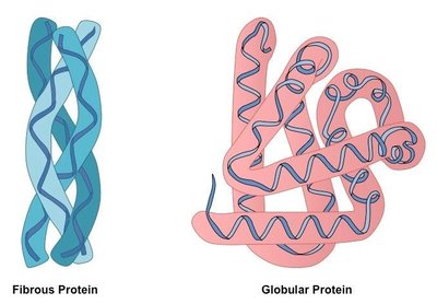
Proteins are essential macromolecules that perform a wide variety of functions in biological systems. They can be classified based on their structure and function into two main types: fibrous and globular proteins.
Fibrous Proteins: These proteins have elongated, fiber-like shapes and provide structural support to cells and tissues. Examples include:
Collagen: Composed of long peptide chains woven together to form strong fibers, found in connective tissues.
Keratin: Structural protein found in scales, horns, wool, nails, and feathers.
Silk: Produced by insects such as spiders and silkworms.
Globular Proteins: These proteins are more compact and spherical in shape. They include enzymes, antibodies, and the subunits of microtubules. Their functions are diverse, including catalysis, transport, and immune defense.
Sickle Cell Disease: Protein Structure and Function
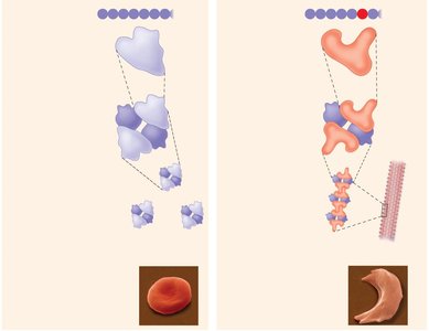
The function of a protein is directly related to its structure. Sickle cell disease is a classic example of how a single amino acid change can alter protein structure and function, leading to disease.
Normal Hemoglobin: Has a specific primary structure (sequence of amino acids) that allows it to carry oxygen efficiently.
Sickle Cell Hemoglobin: A mutation replaces glutamic acid with valine at the sixth position of the beta chain, causing hemoglobin molecules to stick together and form fibers. This distorts red blood cells into a sickle shape, reducing their oxygen-carrying capacity and causing blockages in blood vessels.
Protein Folding and Chaperonins
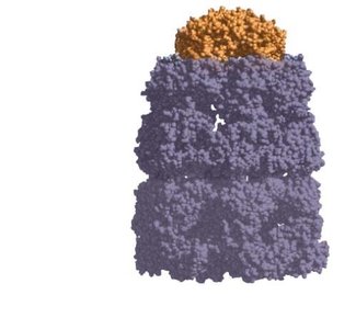
Protein folding is a complex process that determines the final three-dimensional structure of a protein. Proper folding is essential for protein function and is assisted by specialized proteins called chaperonins.
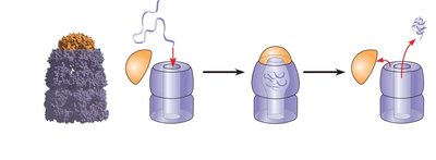
Chaperonins: Do not dictate the final structure but provide a protected environment for polypeptides to fold correctly, preventing misfolding and aggregation.
Folding Process: The unfolded polypeptide enters the chaperonin's hollow cylinder, the cap attaches, creating a hydrophilic environment, and after folding, the cap is released, and the properly folded protein exits.
Nucleic Acids: DNA and RNA
Types and Structure of Nucleic Acids
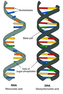
Nucleic acids are polymers made of nucleotide monomers. There are two main types: deoxyribonucleic acid (DNA) and ribonucleic acid (RNA).
DNA: Stores genetic information and is typically double-stranded.
RNA: Involved in protein synthesis and gene regulation; usually single-stranded.
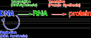
The Central Dogma of Biology
The central dogma describes the flow of genetic information within a biological system:
DNA → RNA → Protein
Transcription: DNA is transcribed into messenger RNA (mRNA) in the nucleus.
Translation: mRNA is translated into a polypeptide (protein) in the cytoplasm.
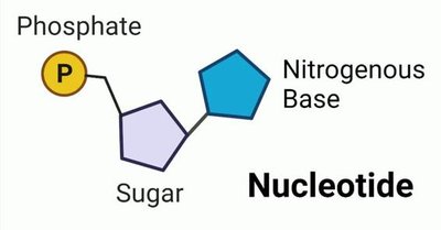
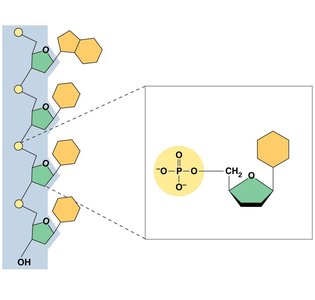
Components of Nucleic Acids
Each nucleotide consists of three components:
Nitrogenous Base: Purines (adenine, guanine) and pyrimidines (cytosine, thymine in DNA; uracil in RNA).
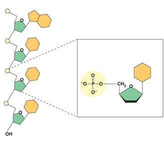
Pentose Sugar: Deoxyribose in DNA, ribose in RNA.
Phosphate Group: Links nucleotides together via phosphodiester bonds.
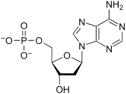
Nucleoside vs Nucleotide
Nucleoside: Consists of a nitrogenous base and a sugar.
Nucleotide: Consists of a nitrogenous base, a sugar, and one or more phosphate groups.
DNA vs RNA: Key Differences
DNA and RNA differ in several important ways:
Sugar: DNA contains deoxyribose; RNA contains ribose.
Nitrogenous Bases: DNA uses adenine (A), guanine (G), cytosine (C), and thymine (T). RNA uses adenine (A), guanine (G), cytosine (C), and uracil (U).
Structure: DNA is typically double-stranded; RNA is usually single-stranded.
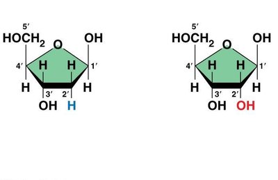
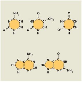
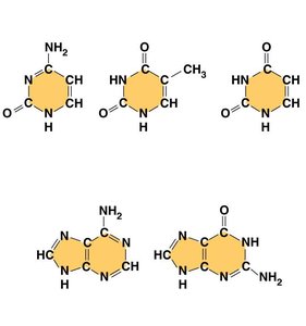
Table: Comparison of DNA and RNA
Feature | DNA | RNA |
|---|---|---|
Sugar | Deoxyribose | Ribose |
Nitrogenous Bases | A, G, C, T | A, G, C, U |
Strands | Double-stranded | Single-stranded |
Function | Genetic information storage | Protein synthesis, gene regulation |
Nitrogenous Bases: Purines and Pyrimidines
Purines: Adenine (A) and Guanine (G); have a double-ring structure.
Pyrimidines: Cytosine (C), Thymine (T, in DNA), and Uracil (U, in RNA); have a single-ring structure.
Base Pairing: In DNA, A pairs with T, and G pairs with C. In RNA, A pairs with U.
Summary
Proteins and nucleic acids are essential macromolecules with diverse structures and functions.
Protein structure determines function, and misfolding can lead to diseases such as sickle cell anemia.
Nucleic acids store and transmit genetic information, with DNA and RNA differing in sugar, bases, and structure.
