 Back
BackBio 100 Lec Chapter 5 Part 2
Study Guide - Smart Notes
 Tailored notes based on your materials, expanded with key definitions, examples, and context.
Bio 100 Lec Chapter 5 (Part 2)
The Structure and Function of Large Biological Molecules
Introduction
Large biological molecules, or macromolecules, are essential for life and include lipids and proteins. Their structures are intricately related to their functions in cells. This guide explores the properties, types, and biological roles of lipids and proteins, focusing on their chemical diversity and importance in cellular processes.
Lipids
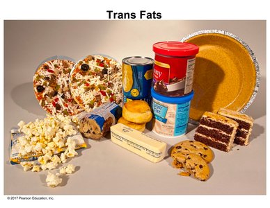
Trans Fats and Saturated Fats
Lipids are a diverse group of hydrophobic molecules, including fats, phospholipids, and steroids. Trans fats are a type of unsaturated fat with trans double bonds, often produced by industrial processing. Unlike natural cis unsaturated fats, trans fats have hydrogen atoms on opposite sides of the double bond, resulting in a more compact structure. This compactness increases shelf life and is desirable in processed foods, but trans fats are linked to increased risk of cardiovascular disease due to their role in plaque formation in arteries.
Saturated fats have no double bonds and pack closely together, contributing to plaque deposits in blood vessels.
Trans fats are even more strongly associated with cardiovascular disease than saturated fats.
Cis unsaturated fats have kinks due to hydrogens on the same side of the double bond, preventing tight packing.
Example: Processed foods such as margarine, baked goods, and fried foods often contain trans fats.
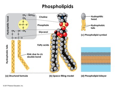
Phospholipids
Phospholipids are major components of cell membranes. Each molecule consists of a glycerol backbone, two fatty acid tails, and a phosphate group attached to a small polar molecule (e.g., choline). The structure is amphipathic, with a hydrophilic (polar) head and hydrophobic (nonpolar) tails. In aqueous environments, phospholipids spontaneously form bilayers, with hydrophobic tails facing inward and hydrophilic heads facing outward, creating a selective barrier for cells.
Hydrophilic head: Contains phosphate and polar groups, interacts with water.
Hydrophobic tails: Composed of hydrocarbons, avoid water.
Bilayer formation: Essential for cell membrane structure and function.
Example: The plasma membrane of cells is a phospholipid bilayer.
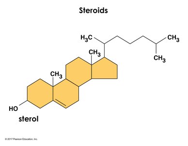
Steroids
Steroids are lipids characterized by a four-ring hydrocarbon skeleton. They include molecules such as cholesterol, testosterone, and estrogen. Steroids with hydroxyl groups are called sterols. Cholesterol is a key component of animal cell membranes and a precursor for other steroids. While essential, cholesterol levels must be regulated, as imbalances are linked to cardiovascular disease.
Structure: Four fused hydrocarbon rings with various functional groups.
Function: Membrane fluidity, hormone synthesis.
Example: Cholesterol in animal cell membranes; testosterone and estrogen as signaling molecules.
Proteins
Diversity of Protein Structure and Function
Proteins are polymers of amino acids and account for more than 50% of the dry mass of most cells. Their diverse structures enable a wide range of functions, including catalysis, defense, storage, transport, signaling, movement, and structural support. The function of a protein is determined by its specific three-dimensional structure.
Enzymes: Catalyze chemical reactions (e.g., DNA polymerase).
Defensive proteins: Protect against disease (e.g., antibodies).
Storage proteins: Store amino acids (e.g., casein in milk).
Transport proteins: Move substances (e.g., hemoglobin).
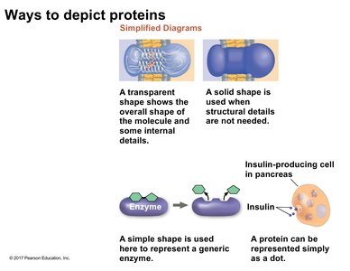
Hormones: Coordinate organismal responses (e.g., insulin).
Receptor proteins: Receive signals from outside the cell.
Motor proteins: Enable movement (e.g., actin, myosin).
Structural proteins: Provide support (e.g., collagen).
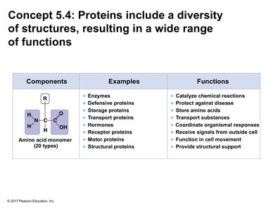
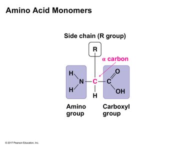
Amino Acid Monomers
Amino acids are the building blocks of proteins. Each amino acid contains a central (alpha) carbon atom bonded to an amino group, a carboxyl group, a hydrogen atom, and a variable side chain (R group). The properties of the R group determine the characteristics and function of each amino acid. At physiological pH, the amino group is typically protonated (NH3+) and the carboxyl group is deprotonated (COO-).
Backbone: Consists of the amino group, alpha carbon, and carboxyl group.
R group (side chain): Confers unique properties to each amino acid.
Classification of Amino Acids
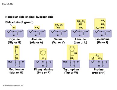
Nonpolar (Hydrophobic) Side Chains
Nonpolar amino acids have side chains that are hydrophobic, often consisting of hydrocarbons. These amino acids tend to cluster in the interior of proteins, away from water, and are abundant in membrane-spanning regions.
Examples: Glycine, Alanine, Valine, Leucine, Isoleucine, Methionine, Phenylalanine, Tryptophan, Proline.
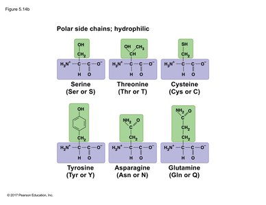
Polar (Hydrophilic) Side Chains
Polar amino acids have side chains that can form hydrogen bonds with water, making them hydrophilic. These amino acids are often found on the surfaces of proteins, interacting with the aqueous environment.
Examples: Serine, Threonine, Cysteine, Tyrosine, Asparagine, Glutamine.
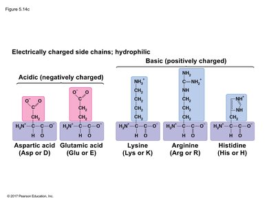
Electrically Charged (Hydrophilic) Side Chains
Some amino acids have side chains that are charged at physiological pH. Acidic side chains are negatively charged, while basic side chains are positively charged. These amino acids are important for protein solubility and function, including enzyme active sites and binding interactions.
Acidic (negatively charged): Aspartic acid, Glutamic acid.
Basic (positively charged): Lysine, Arginine, Histidine.
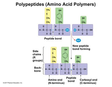
Polypeptides and Peptide Bonds
Proteins are formed by linking amino acids through peptide bonds in a condensation reaction (dehydration synthesis), releasing water. Polypeptides are linear chains with directionality, having an N-terminus (amino end) and a C-terminus (carboxyl end). The sequence of amino acids (primary structure) determines the protein's properties and function.
Peptide bond: Covalent bond between the carboxyl group of one amino acid and the amino group of the next.
Directionality: Synthesized from N-terminus to C-terminus.
Protein Structure and Function
The specific activities of proteins are determined by their three-dimensional architecture. The folding and shape of a protein are critical for its function, and even small changes in structure can have significant effects on activity.
Structure-function relationship: The unique shape of a protein enables its specific function in the cell.
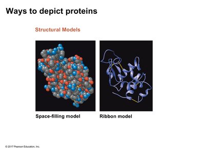
Ways to Depict Proteins
Proteins can be represented using various models, each highlighting different aspects of their structure:
Space-filling model: Shows the spatial arrangement of atoms.
Ribbon model: Emphasizes the folding pattern of the polypeptide backbone.
Simplified diagrams: Used to illustrate overall shape, function, or location without detailed structure.
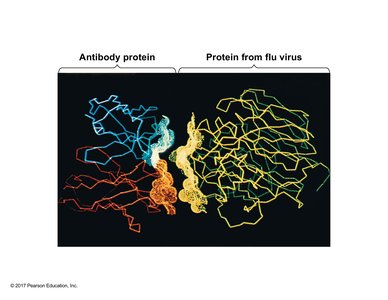
Protein-Protein Interactions
Proteins often interact with other molecules, including other proteins. For example, antibody proteins can specifically bind to foreign proteins (antigens) from pathogens such as viruses, neutralizing them and marking them for destruction by the immune system.
Example: Antibody binding to a flu virus protein demonstrates the specificity and complementarity of protein-protein interactions.
Levels of Protein Structure
Proteins have up to four levels of structure:
Primary structure: Linear sequence of amino acids in a polypeptide chain, determined by peptide bonds. The sequence dictates higher levels of structure and ultimately the protein's function.
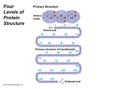
Additional info: Higher levels of protein structure include secondary (alpha helices and beta sheets), tertiary (overall 3D folding), and quaternary (assembly of multiple polypeptides), which together determine the final functional form of the protein.
