 Back
The Structure and Function of the Mammalian Heart: Circulation, Heart Rate, ECG, and Blood Pressure
Study Guide - Smart Notes
 Tailored notes based on your materials, expanded with key definitions, examples, and context.
Structure and Function of the Mammalian Heart
Overview of Heart Circulation
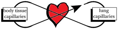
The mammalian heart is a central organ responsible for pumping blood throughout the body in a double circulation system. This system separates pulmonary (lung) and systemic (body) circuits, ensuring efficient oxygen delivery and waste removal.
Double Circulation: Blood passes through the heart twice during each complete circuit—once through the pulmonary circuit (right side) and once through the systemic circuit (left side).
Pulmonary Circuit: Right side of the heart pumps deoxygenated blood to the lungs for oxygenation.
Systemic Circuit: Left side of the heart pumps oxygenated blood to body tissues.
Evolutionary Comparison: Mammals, birds, and crocodiles have a four-chambered heart with double circulation; fish have single circulation; amphibians and reptiles have a three-chambered heart with double low-pressure circulation.
Anatomy of the Heart
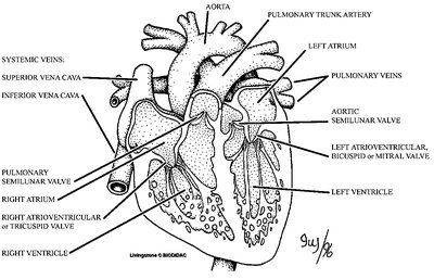
The heart consists of four chambers: two atria (receiving chambers) and two ventricles (pumping chambers). Valves ensure unidirectional blood flow and prevent mixing of oxygenated and deoxygenated blood.
Atria: Right atrium receives deoxygenated blood from the body; left atrium receives oxygenated blood from the lungs.
Ventricles: Right ventricle pumps blood to the lungs; left ventricle pumps blood to the body.
Valves:
Atrioventricular Valves: Tricuspid (right) and bicuspid/mitral (left) valves between atria and ventricles.
Semilunar Valves: Pulmonary (right) and aortic (left) valves at the exits of the ventricles.
Interventricular Septum: Thick tissue separating the ventricles.
Blood Vessels
Blood vessels are classified based on their function and structure:
Arteries: Thick-walled, carry blood away from the heart; largest is the aorta.
Veins: Thin-walled, carry blood toward the heart; largest are the superior and inferior vena cava.
Capillaries: Very thin-walled, facilitate exchange of gases and metabolites.
Pathway of Blood Flow
Blood follows a specific pathway through the heart and body, passing through various chambers and valves.
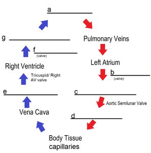
Deoxygenated blood: Body tissues → vena cava → right atrium → tricuspid valve → right ventricle → pulmonary semilunar valve → pulmonary artery → lungs
Oxygenated blood: Lungs → pulmonary veins → left atrium → bicuspid/mitral valve → left ventricle → aortic semilunar valve → aorta → body tissues
Heart Rate and Electrical Activity
Heart Rate
The heart rate is the number of beats per minute, determined by the rhythmic impulses generated in the sinoatrial (SA) node of the right atrium.
Normal adult heart rate: 60–100 beats per minute.
Pulse measurement: Felt in arteries close to the skin (e.g., radial, carotid).
Electrocardiogram (ECG/EKG)
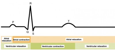
An ECG records the electrical activity of the heart, showing characteristic waves corresponding to different phases of the cardiac cycle.
P-wave: Atrial contraction (depolarization).
QRS complex: Ventricular contraction (depolarization) and atrial relaxation (repolarization).
T-wave: Ventricular relaxation (repolarization).
Systole: Chamber contraction.
Diastole: Chamber relaxation.
Blood Pressure
Measurement and Significance
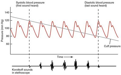
Blood pressure is the force exerted by blood on arterial walls, measured in millimeters of mercury (mmHg). It fluctuates between systolic (peak) and diastolic (low) values due to the pumping action of the ventricles.
Systolic Pressure: Highest pressure during ventricular contraction.
Diastolic Pressure: Lowest pressure during ventricular relaxation.
Measurement: Sphygmomanometer (blood pressure cuff) and stethoscope are used to detect Korotkoff sounds.
Korotkoff Sounds: Rhythmic tapping sounds heard as cuff pressure falls between systolic and diastolic values.
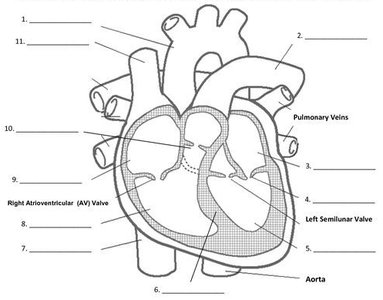
Heart Structure and Blood Flow Worksheet
Labeling the Heart
Students are required to label the heart diagram and indicate the direction of oxygenated (red) and deoxygenated (blue) blood flow.
Double Circulation Flow of Blood
Students fill out a diagram showing the flow of blood through pulmonary and systemic circuits, identifying key vessels and valves.
Key Terms and Concepts
Heart Murmur: Abnormal sound caused by valve defects or leaks.
Sinoatrial Node: Pacemaker of the heart, sets heart rate.
Systole: Contraction phase of the heart.
Diastole: Relaxation phase of the heart.
Korotkoff Sounds: Sounds heard during blood pressure measurement.
Sample Table: Comparison of Circulatory Systems
The following table compares the circulatory systems of different vertebrate groups:
Group | Heart Chambers | Circulation Type | Efficiency |
|---|---|---|---|
Fish | 2 (1 atrium, 1 ventricle) | Single | Low |
Amphibians/Reptiles | 3 (2 atria, 1 ventricle) | Double (low pressure) | Moderate |
Mammals/Birds/Crocodiles | 4 (2 atria, 2 ventricles) | Double (high pressure) | High |
Key Equations
Blood Pressure:
Heart Rate Calculation:
Summary of Lab Procedures
Measure heart rate by palpating an artery and counting beats.
Record ECG using electrodes placed on wrists and ankles.
Measure blood pressure using a sphygmomanometer and stethoscope, noting systolic and diastolic values.
Review Questions
Through which vessels does blood arrive at the right atrium? Answer: Superior and inferior vena cava.
Where does blood in the pulmonary artery travel to next? Answer: Lungs.
Which chamber pumps blood to the pulmonary artery? Answer: Right ventricle.
Is blood on the right side of the heart oxygenated or deoxygenated? Answer: Deoxygenated.
Which vessels carry blood back to the left side of the heart? Answer: Pulmonary veins.
Which valve guards the entrance to the left ventricle? Answer: Bicuspid/mitral valve.
Blood from the left atrium enters which chamber? Answer: Left ventricle.
Which chamber (include right or left) is three times thicker than the other? Answer: Left ventricle.
Are systemic arteries connected to the heart's right or left side? Answer: Left side.
What is the body’s largest artery? Answer: Aorta.
Which valves are shutting when you hear “lubb”? Answer: Atrioventricular valves.
A minor heart valve leak causes a sound called a heart murmur.
Your heart rate is set by rhythmic impulses generated in the sinoatrial node.
Is chamber contraction called systole or diastole? Answer: Systole.
How many chambers does a frog (amphibian) heart have? Answer: Three.
During which part of the ECG do the atria relax? Answer: QRS complex.
During which part of the ECG do the ventricles contract? Answer: QRS complex.
Vessels that carry blood towards the heart are always called veins.
Blood pressure is measured in a/an artery.
Is systolic pressure when the Korotkoff sounds begin or cease? Answer: Begin.
When the pressure in a sphygmomanometer is higher than systolic pressure, you hear no sound (silent).
Additional info: Academic context was added to clarify evolutionary comparisons, ECG interpretation, and blood pressure measurement.
