 Back
The Urinary System: Structure, Function, and Physiology
Study Guide - Smart Notes
 Tailored notes based on your materials, expanded with key definitions, examples, and context.
The Urinary System
Overview and Major Functions
The urinary system is essential for maintaining homeostasis by regulating the composition and volume of blood, removing metabolic wastes, and balancing electrolytes and pH. It consists of the kidneys, ureters, urinary bladder, and urethra.
Regulation of water and solute concentration: Maintains total water volume and solute concentration in the body.
Ion balance: Controls extracellular fluid (ECF) ion concentrations.
Acid-base balance: Ensures long-term regulation of blood pH.
Excretion: Removes metabolic wastes, toxins, and drugs from the body.
Endocrine functions: Secretes renin (regulates blood pressure), erythropoietin (stimulates red blood cell production), and activates vitamin D.
Gluconeogenesis: Generates glucose during prolonged fasting.
Organs of the Urinary System
Kidneys: Major excretory organs that filter blood and form urine.
Ureters: Muscular tubes that transport urine from the kidneys to the urinary bladder.
Urinary bladder: Temporary storage reservoir for urine.
Urethra: Tube that conveys urine from the bladder to the outside of the body.
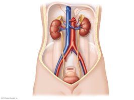
Anatomy of the Kidneys
Location and Structure
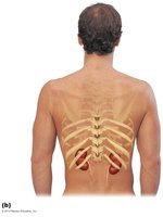
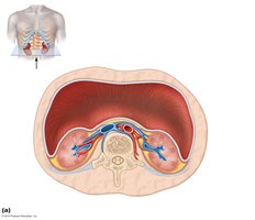
The kidneys are retroperitoneal organs located against the posterior body wall, protected by the lower ribs and surrounded by supportive tissue layers.
Renal fascia: Outermost layer anchoring the kidney.
Perirenal fat capsule: Cushions and protects the kidney.
Fibrous capsule: Prevents infection and trauma.
Internal Anatomy
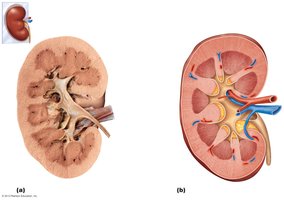
Each kidney has a distinct internal structure composed of the cortex, medulla, and renal pelvis.
Renal cortex: Outer region containing renal corpuscles and convoluted tubules.
Renal medulla: Inner region with renal pyramids and columns.
Renal pelvis: Funnel-shaped structure that collects urine and channels it to the ureter.
Calyces: Minor and major calyces collect urine from the papillae of the pyramids.
Homeostatic Imbalances
Pyelitis: Infection of the renal pelvis and calyces.
Pyelonephritis: Infection/inflammation of the entire kidney, usually treatable with antibiotics.
Nephrons: The Functional Units of the Kidney
Structure of the Nephron
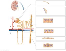
Nephrons are the microscopic structural and functional units of the kidney responsible for urine formation. Each nephron consists of a renal corpuscle and a renal tubule.
Renal corpuscle: Includes the glomerulus (a tuft of capillaries) and the glomerular (Bowman's) capsule.
Renal tubule: Composed of the proximal convoluted tubule (PCT), nephron loop (loop of Henle), and distal convoluted tubule (DCT).
Collecting duct: Receives filtrate from multiple nephrons and channels it toward the renal pelvis.
Classes of Nephrons
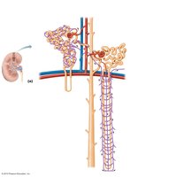
Cortical nephrons: 85% of nephrons; located almost entirely in the cortex.
Juxtamedullary nephrons: Long nephron loops extend deep into the medulla; essential for producing concentrated urine.
Nephron Capillary Beds
Peritubular capillaries: Surround cortical nephrons; adapted for absorption of water and solutes.
Vasa recta: Long, straight capillaries paralleling the nephron loops of juxtamedullary nephrons; crucial for forming concentrated urine.
Mechanisms of Urine Formation
Overview of Renal Processes
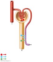
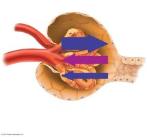
The kidneys process about 180 liters of fluid daily, but only about 1.5 liters are excreted as urine. Three main processes adjust plasma composition:
Glomerular filtration: Passive process producing a cell- and protein-free filtrate.
Tubular reabsorption: Selectively returns 99% of substances from filtrate to blood.
Tubular secretion: Selectively moves substances from blood to filtrate.
Glomerular Filtration
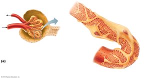
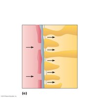
Filtration occurs in the renal corpuscle, driven by hydrostatic pressure. The filtration membrane consists of three layers:
Capillary endothelium
Basement membrane
Foot processes of podocytes (visceral layer of Bowman's capsule)
Net Filtration Pressure (NFP)
The net filtration pressure determines the rate of filtrate formation:
Formula:
Typical values: mm Hg
HPgc: Glomerular capillary hydrostatic pressure
HPcs: Capsular hydrostatic pressure
OPgc: Glomerular capillary oncotic pressure
Tubular Reabsorption
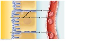
Most filtrate is reabsorbed into the blood via selective transepithelial transport. Reabsorption can occur via two routes:
Transcellular route: Through the tubule cells.
Paracellular route: Between tubule cells, especially in the proximal convoluted tubule (PCT).
Mechanisms of Reabsorption
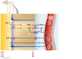
Primary active transport: Na+ is pumped out of the tubule cell by Na+-K+ ATPase at the basolateral membrane.
Secondary active transport: Glucose, amino acids, and other nutrients are co-transported with Na+.
Osmosis: Water follows solute reabsorption via aquaporins.
Passive diffusion: Lipid-soluble substances and some ions move down their gradients.
Transport Maximum (Tm)
Each substance has a transport maximum, reflecting the number of available carriers. Excess amounts are excreted in urine (e.g., glucose in diabetes mellitus).
Reabsorptive Capabilities by Region
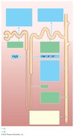
PCT: Site of most reabsorption (all nutrients, 65% of Na+ and water, many ions).
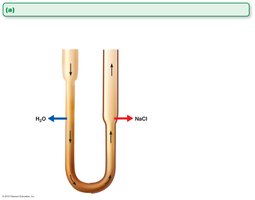
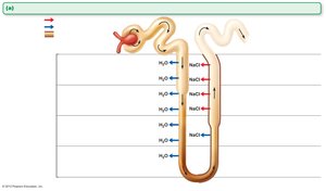
Nephron loop: Descending limb is permeable to water, ascending limb to solutes (Na+, Cl-).
DCT and collecting duct: Reabsorption is hormonally regulated (aldosterone, ADH, parathyroid hormone).
Tubular Secretion
Tubular secretion removes substances from blood into the filtrate, including drugs, excess K+, and H+ to regulate pH. Most secretion occurs in the PCT.
Regulation of Urine Concentration and Volume
Osmolality and Countercurrent Mechanism
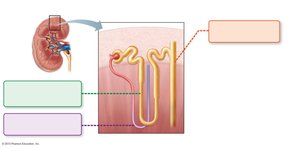
The kidneys maintain plasma osmolality (~300 mOsm) by regulating urine concentration and volume using the countercurrent mechanism. This mechanism establishes and maintains an osmotic gradient from cortex to medulla (300–1200 mOsm), allowing the kidneys to produce urine of varying concentration.
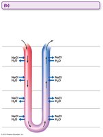
Role of Antidiuretic Hormone (ADH)
ADH regulates the number of aquaporins in the collecting duct, controlling water reabsorption and thus urine concentration. Overhydration leads to low ADH and dilute urine; dehydration leads to high ADH and concentrated urine.
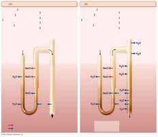
Diuretics
ADH inhibitors: Alcohol increases urine output.
Na+ reabsorption inhibitors: Caffeine and some drugs increase urine output by inhibiting sodium reabsorption.
Loop diuretics: Inhibit medullary gradient formation.
Osmotic diuretics: Substances not reabsorbed (e.g., glucose in diabetes) keep water in urine.
Physical and Chemical Characteristics of Urine
Physical Characteristics
Color and transparency: Clear, pale to deep yellow (urochrome pigment); abnormal colors may indicate pathology or diet.
Odor: Slightly aromatic when fresh; may develop ammonia odor upon standing or be altered by certain foods and drugs.
pH: Slightly acidic (pH ~6); varies with diet and health.
Specific gravity: 1.001 to 1.035, depending on solute concentration.
Chemical Composition
Water: 95% of urine volume.
Solutes: 5%, including nitrogenous wastes (urea, uric acid, creatinine) and ions (Na+, K+, PO43–, SO42–, Ca2+, Mg2+, HCO3–).
Abnormal components: High concentrations of any constituent, or presence of blood proteins, WBCs, or bile pigments, may indicate disease.
Clinical Considerations
Homeostatic Imbalances
Chronic renal disease: GFR < 60 ml/min for 3 months (e.g., diabetes, hypertension).
Renal failure: GFR < 15 ml/min; leads to uremia, requiring dialysis or transplant.
