 Back
The Urinary System: Structure, Function, and Regulation
Study Guide - Smart Notes
 Tailored notes based on your materials, expanded with key definitions, examples, and context.
The Urinary System
Overview and Role in Excretion
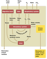
The urinary system is essential for maintaining homeostasis by regulating the composition and volume of body fluids. It removes metabolic wastes, excess solutes, and water from the body, working in concert with other organ systems involved in excretion.
Excretion: The process of removing wastes and excess materials from the body.
Participating organ systems:
Digestive system: Eliminates food residues and liver-produced wastes.
Respiratory system: Removes carbon dioxide.
Integumentary system: Excretes water and salts via sweat.
Urinary system: Excretes nitrogenous wastes, excess solutes, and water.
Major Structures of the Urinary System
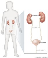
The urinary system consists of paired kidneys, ureters, a urinary bladder, and a urethra. Each structure plays a specific role in urine formation, storage, and elimination.
Kidneys: Produce urine by filtering blood and regulating water and solute balance.
Ureters: Muscular tubes that transport urine from the kidneys to the bladder.
Urinary bladder: Stores urine until excretion (capacity: 600–1000 mL).
Urethra: Conducts urine from the bladder to the outside of the body.
Kidney Structure and Function
Gross Anatomy of the Kidney
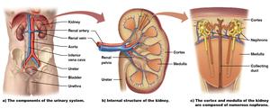
The kidney is a bean-shaped organ with distinct regions that contribute to urine formation and transport.
Cortex: The outer region where urine is produced.
Medulla: The inner region that transports urine toward the renal pelvis.
Renal pelvis: A hollow space where urine collects before entering the ureter.
Microscopic Structure: The Nephron
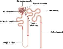
The nephron is the functional unit of the kidney, responsible for filtering blood and forming urine. Each kidney contains about one million nephrons.
Glomerulus: A ball of capillaries where filtration occurs.
Tubule: A thin, hollow tube of epithelial cells with four main regions:
Proximal tubule
Loop of Henle
Distal tubule
Collecting duct
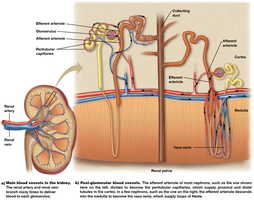
Blood Supply to the Nephron
Specialized blood vessels supply and drain the nephron, facilitating filtration and reabsorption.
Renal artery: Supplies blood to the kidney.
Glomerular capillaries: Site of filtration.
Renal vein: Returns filtered blood to circulation.
Urine Formation
Three Main Processes
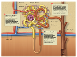
Urine formation involves three sequential processes:
Glomerular filtration: Movement of protein-free plasma from blood into the glomerular capsule, driven by high blood pressure.
Tubular reabsorption: Return of most filtered water and solutes (e.g., glucose, amino acids, bicarbonate) to the blood, primarily in the proximal tubule.
Tubular secretion: Addition of specific solutes (e.g., drugs, H+, K+) from blood into the tubule for excretion.
Filtration and Reabsorption Details
Filtration: Highly selective, excludes proteins and blood cells. Regulated by arteriole diameter and sympathetic nervous system activity.
Reabsorption:
100% of glucose, amino acids, and bicarbonate are reabsorbed.
50% of urea is reabsorbed.
Sodium is actively transported, creating gradients for water and other solutes to follow.
Water moves via aquaporins (water channels).
Tubular Secretion
Secretion helps regulate chemical levels and remove harmful substances from the body.
Substances secreted include: penicillin, drugs, pesticides, hydrogen ions, ammonium, potassium.
Regulation of Water and Solute Balance
Water Balance and ADH
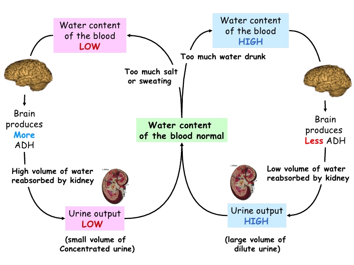
The kidneys adjust water output to maintain homeostasis. Antidiuretic hormone (ADH) from the posterior pituitary increases water reabsorption in response to dehydration or high blood osmolarity.
High water intake: Less ADH, more dilute urine.
Low water intake or dehydration: More ADH, concentrated urine.
Salt Balance and Aldosterone
Aldosterone, an adrenal hormone, increases sodium reabsorption in the distal tubule and collecting duct. The renin-angiotensin system regulates aldosterone release in response to low blood volume or pressure.
Renin-angiotensin system:
Renin (from kidney) triggers production of angiotensin II.
Angiotensin II causes vasoconstriction and stimulates aldosterone release.
Aldosterone increases sodium (and water) reabsorption, raising blood volume and pressure.
Atrial Natriuretic Hormone (ANH)
ANH is released by the heart's atria in response to high blood volume. It inhibits sodium reabsorption, increasing sodium and water excretion to reduce blood volume.
Acid-Base Balance and Other Kidney Functions
Maintaining Blood pH
The kidneys help maintain blood pH (7.35–7.45) by reabsorbing bicarbonate and excreting hydrogen ions as ammonium.
Erythropoietin and Vitamin D Activation
Erythropoietin: Hormone released by the kidney in response to low oxygen, stimulating red blood cell production in bone marrow.
Vitamin D activation: Kidneys convert inactive vitamin D to its active form, essential for calcium homeostasis.
Summary Table: Key Hormones in Kidney Regulation
Hormone | Source | Main Effect |
|---|---|---|
ADH (Antidiuretic Hormone) | Posterior pituitary | Increases water reabsorption in collecting ducts |
Aldosterone | Adrenal cortex | Increases sodium (and water) reabsorption |
ANH (Atrial Natriuretic Hormone) | Atria of heart | Inhibits sodium reabsorption, increases excretion |
Erythropoietin | Kidney | Stimulates red blood cell production |
Clinical Connections
Diuretics: Substances that increase urine formation and excretion, used to treat hypertension and edema.
ACE inhibitors: Medications that block angiotensin II formation, lowering blood pressure by reducing vasoconstriction and aldosterone secretion.
