 Back
BackTissue Histology and Comparative Vertebrate Anatomy: Study Guide
Study Guide - Smart Notes
 Tailored notes based on your materials, expanded with key definitions, examples, and context.
Laboratory 8: Tissue Histology
Introduction to Tissues
Tissues are groups of cells with a specific function in the body. In animals, there are four basic types of tissues: epithelial, connective, muscular, and nervous. Each tissue type is specialized for particular roles and forms the foundation for organ structure and function. Organs are composed of multiple tissue types working together to perform complex biological functions.
Organ System | Main Function | Principal Organs |
|---|---|---|
Integumentary | Protection, temperature regulation | Skin, hair, nails |
Muscular | Movement, heat production | Skeletal muscles |
Skeletal | Support, protection, blood formation | Bones, cartilage |
Nervous | Coordination, control | Brain, spinal cord, nerves |
Cardiovascular | Transport of substances | Heart, blood vessels |
Respiratory | Gas exchange | Lungs, trachea |
Digestive | Breakdown and absorption of food | Stomach, intestines |
Urinary | Excretion of wastes | Kidneys, bladder |
Reproductive | Production of offspring | Ovaries, testes |
Objectives
Examine the structure and function of the four tissue types in animals.
Identify tissues under the microscope and relate structure to function.
Describe the relationship between tissues, organs, and organ systems.
A. Epithelial Tissue
Structure and Function
Epithelial tissue covers surfaces, lines cavities, and forms glands. It is composed of tightly packed cells with minimal extracellular material. Epithelial cells have polarity, with an apical surface (exposed to the exterior or cavity) and a basal surface (attached to underlying connective tissue via a basement membrane).
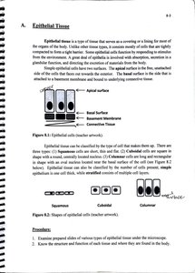
Simple epithelium: Single cell layer; specialized for absorption, secretion, and filtration.
Stratified epithelium: Multiple cell layers; provides protection in areas of abrasion.
Cell shapes include squamous (flat), cuboidal (cube-shaped), and columnar (tall, column-like).
Types of Epithelial Tissue
Simple Squamous: Single layer of flat cells; found in lungs, blood vessels (for diffusion/filtration).
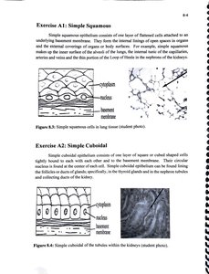
Simple Cuboidal: Single layer of cube-shaped cells; found in kidney tubules, glands (for secretion/absorption).
Simple Columnar: Single layer of tall cells; lines digestive tract (for absorption/secretion).
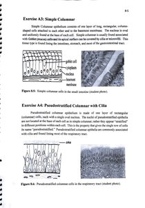
Pseudostratified Columnar (with cilia): Appears layered but all cells touch the basement membrane; lines respiratory tract (for secretion, movement of mucus).
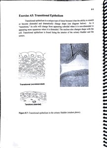
Transitional Epithelium: Multiple layers; cells change shape when stretched; found in urinary bladder.
Stratified Squamous: Many layers of flat cells; found in skin, mouth, esophagus (for protection).
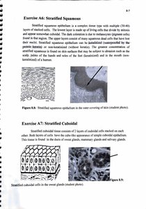
Stratified Cuboidal: Two or more layers of cube-shaped cells; found in ducts of sweat glands, mammary glands.
B. Connective Tissue
Structure and Function
Connective tissue is the most abundant tissue type in the body, providing support, binding, and protection. It consists of cells embedded in an extracellular matrix of fibers and ground substance. The composition and arrangement of these components determine the tissue's properties.
Loose Connective Tissue: Areolar, adipose, reticular
Dense Connective Tissue: Regular, irregular, elastic
Cartilage: Hyaline, elastic, fibrocartilage
Other: Blood, bone (osseous tissue)
Loose Connective Tissue
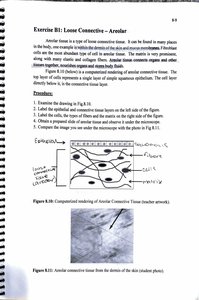
Areolar: Supports and binds other tissues; found under epithelia.
Adipose: Stores fat; insulates and protects organs.
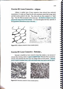
Reticular: Forms a soft internal skeleton for lymphoid organs.
Dense Connective Tissue
Dense Irregular: Collagen fibers in multiple directions; found in dermis.
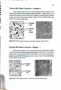
Dense Regular: Collagen fibers aligned; found in tendons and ligaments.
Elastic: Contains elastic fibers; found in ligaments and arteries.
Cartilage
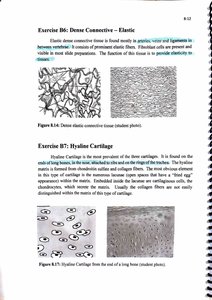
Hyaline: Most common; supports and reinforces; found in nose, trachea, ends of long bones.
Elastic: Flexible; found in ear, epiglottis.
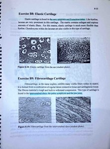
Fibrocartilage: Strongest; resists compression; found in intervertebral discs, pubic symphysis.
Other Connective Tissues
Blood: Cells suspended in plasma; transports gases, nutrients, wastes.

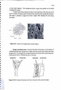
Osseous (Bone) Tissue: Supports, protects, stores minerals; compact and spongy types.
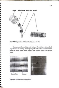
C. Muscular Tissue
Types and Functions
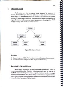
Muscular tissue is specialized for contraction and movement. There are three types:
Skeletal Muscle: Voluntary, striated, multinucleated; attached to bones for movement.
Cardiac Muscle: Involuntary, striated, branched; found only in the heart.
Smooth Muscle: Involuntary, non-striated, spindle-shaped; found in walls of hollow organs.
Microscopic Structure
Skeletal muscle fibers are organized into bundles called fascicles. Each fiber contains myofibrils responsible for contraction.
Cardiac muscle cells are branched and connected by intercalated discs for synchronized contraction.
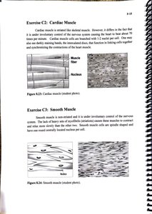
Smooth muscle cells are spindle-shaped with a single nucleus; found in the digestive tract, blood vessels, etc.
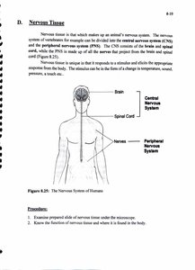
D. Nervous Tissue
Structure and Function
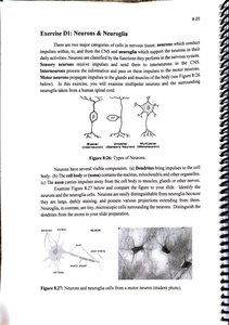
Nervous tissue is specialized for communication via electrical impulses. It consists of two main cell types: neurons (transmit signals) and neuroglia (support and protect neurons). The nervous system is divided into the central nervous system (CNS) and peripheral nervous system (PNS).
Neurons: Large cells with dendrites (receive signals), a cell body, and an axon (transmits signals).
Neuroglia: Smaller supporting cells; maintain homeostasis, form myelin, and provide support and protection.
Laboratory 9: Comparative Vertebrate Anatomy – External
Introduction
Comparative vertebrate anatomy examines the similarities and differences in the external and internal structures of vertebrates. This field provides insight into evolutionary relationships and adaptations to different environments.
Objectives
Examine the external anatomy of four vertebrate animals: shark, frog, turtle, and rat.
Identify major anatomical features and compare adaptations among these species.
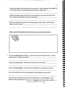
Vertebrate Fish – Dogfish Shark (Squalus acanthias)
The dogfish shark is a cartilaginous fish with adaptations for aquatic life, including fins, gill slits, and placoid scales. External features include the mouth, nostrils, eyes, gill slits, fins, and lateral line system.
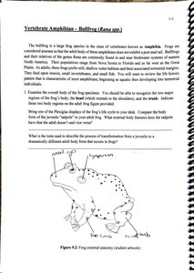
Vertebrate Amphibian – Bullfrog (Rana catesbeiana)
Bullfrogs are amphibians with moist skin, webbed feet, and adaptations for both aquatic and terrestrial environments. Key external features include the head, trunk, limbs, tympanic membrane, and cloacal opening.
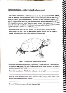
Vertebrate Reptile – Slider Turtle (Trachemys scripta)
The slider turtle is a freshwater reptile with a hard shell (carapace and plastron), scaly skin, and limbs adapted for swimming and walking. External features include the beak, limbs, claws, and tail.
Review Questions and Comparative Tables
Review questions test understanding of tissue types, their functions, and anatomical adaptations in vertebrates.
Muscle Type | Nuclei | Striated | Cell Shape | Control | Location |
|---|---|---|---|---|---|
Skeletal | Multi | Yes | Long, cylindrical | Voluntary | Attached to bones |
Cardiac | 1-2 | Yes | Branched | Involuntary | Heart |
Smooth | 1 | No | Spindle-shaped | Involuntary | Walls of hollow organs |
Example: Blood is classified as a connective tissue because its cells are suspended in plasma, a liquid matrix, allowing for transport of substances throughout the body.
