 Back
Unit 1: Cell Biology Study Guide – Biomolecules, Cell Types, and Viruses
Study Guide - Smart Notes
 Tailored notes based on your materials, expanded with key definitions, examples, and context.
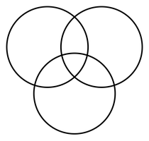
Q1. Fill in the chart with functions and examples for each biomolecule: Carbohydrates, Lipids, Proteins
Background
Topic: Biomolecules and Their Functions
This question tests your understanding of the main biomolecules found in cells, their functions, and examples of each.
Key Terms:
Carbohydrates: Molecules that provide quick energy and structural support.
Lipids: Molecules that store long-term energy and make up cell membranes.
Proteins: Molecules that build body structures, speed up reactions (enzymes), and help with healing.
Step-by-Step Guidance
Start by listing the main function of each biomolecule (e.g., energy, structure, catalysis).
Think of examples for each: for carbohydrates, consider glucose or starch; for lipids, think of fats or oils; for proteins, consider enzymes or muscle fibers.
Compare and contrast their roles: Which provide energy quickly? Which are for long-term energy? Which help build or repair tissues?
Fill in the chart with these functions and examples, making sure to note any overlap (e.g., both carbs and lipids provide energy).
Try solving on your own before revealing the answer!
Final Answer:
Carbohydrates: Quick energy (glucose, starch); Lipids: Long-term energy, cell membrane (fats, oils); Proteins: Structure, enzymes, healing (muscle, enzymes).
Each biomolecule has unique and overlapping functions in the cell.
Q2. Write a cheat sheet for Johnny: Which biomolecules are best for sprinting, weight lifting, and marathon training?
Background
Topic: Biomolecules and Exercise
This question tests your ability to connect biomolecule functions to physical activities and energy needs.
Key Terms:
Sprinting: Requires quick energy (carbohydrates).
Weight lifting: Needs muscle building and repair (proteins), plus energy (carbs, lipids).
Marathon training: Needs sustained energy (lipids), muscle endurance (proteins).
Step-by-Step Guidance
Identify the energy demands of each activity: short bursts vs. sustained effort.
Match the biomolecule to the activity: carbs for quick energy, lipids for long-term, proteins for muscle repair.
Consider how each biomolecule supports recovery and performance.
Summarize which biomolecules are most important for each type of exercise.
Try solving on your own before revealing the answer!
Final Answer:
Sprinting: Carbs and proteins; Weight lifting: Proteins, carbs, lipids; Marathon: Lipids and proteins.
Each activity uses biomolecules differently based on energy and muscle needs.
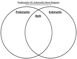
Q3. Compare prokaryotic and eukaryotic cells using a Venn diagram.
Background
Topic: Cell Types
This question tests your understanding of the differences and similarities between prokaryotic and eukaryotic cells.
Key Terms:
Prokaryotic: No membrane-bound organelles, DNA in cytoplasm, cell wall.
Eukaryotic: Membrane-bound organelles (nucleus, mitochondria, chloroplasts), DNA in nucleus.
Both: Cell membrane, ribosomes, cytoplasm, DNA.
Step-by-Step Guidance
List features unique to prokaryotes (e.g., cell wall, no nucleus).
List features unique to eukaryotes (e.g., nucleus, mitochondria).
Identify features shared by both (e.g., DNA, ribosomes).
Organize these features in a Venn diagram format.
Try solving on your own before revealing the answer!
Final Answer:
Prokaryotic: No nucleus, no membrane-bound organelles; Eukaryotic: Nucleus, organelles; Both: DNA, cell membrane, ribosomes.
The Venn diagram helps visualize similarities and differences.
Q4. What two organelles are thought to be a result of endosymbiosis?
Background
Topic: Endosymbiotic Theory
This question tests your knowledge of cell evolution and organelle origins.
Key Terms:
Endosymbiosis: One organism lives inside another, leading to organelle formation.
Mitochondria and chloroplasts: Organelles believed to have originated from endosymbiotic events.
Step-by-Step Guidance
Recall which organelles have their own DNA and double membranes.
Think about which organelles are involved in energy production (ATP, photosynthesis).
Connect these features to the endosymbiotic theory.
Try solving on your own before revealing the answer!
Final Answer:
Mitochondria and chloroplasts are thought to be the result of endosymbiosis.
They have their own DNA and double membranes, supporting this theory.
Q5. Define endosymbiosis.
Background
Topic: Cell Evolution
This question tests your understanding of the process by which certain organelles originated.
Key Terms:
Endosymbiosis: A symbiotic relationship where one organism lives inside another.
Step-by-Step Guidance
Think about the meaning of 'endo' (inside) and 'symbiosis' (living together).
Consider how this process could lead to organelle formation.
Try solving on your own before revealing the answer!
Final Answer:
Endosymbiosis is when one organism lives inside the cells of another organism.
This process explains the origin of mitochondria and chloroplasts.
Q6. What features are shared by all types of cells?
Background
Topic: Universal Cell Features
This question tests your understanding of the basic components found in all cells.
Key Terms:
DNA: Genetic material present in all cells.
Cell membrane: Boundary of the cell.
Ribosomes: Protein synthesis machinery.
Cytoplasm: Internal fluid of the cell.
Step-by-Step Guidance
List the structures found in both prokaryotic and eukaryotic cells.
Consider the functions of these structures.
Try solving on your own before revealing the answer!
Final Answer:
All cells share DNA, cell membrane, ribosomes, and cytoplasm.
These are essential for cell function and survival.
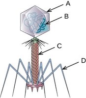
Q7. Label the following parts of a bacteriophage: A, B, C, D
Background
Topic: Virus Structure
This question tests your ability to identify the structural components of a bacteriophage.
Key Terms:
Capsid head: Protein shell containing genetic material.
RNA/DNA: Genetic material inside the capsid.
Sheath: Structure used for injecting genetic material.
Tail fibers: Used for attachment to host cells.
Step-by-Step Guidance
Identify the capsid head (A) at the top of the bacteriophage.
Locate the genetic material (B) inside the capsid.
Find the sheath (C) below the capsid.
Point out the tail fibers (D) at the bottom, used for attachment.
Try solving on your own before revealing the answer!
Final Answer:
A: Capsid head; B: RNA/DNA; C: Sheath; D: Tail fibers.
Each part plays a role in infection and attachment.
Q8. Which part of the bacteriophage attaches to the host cell?
Background
Topic: Virus-Host Interaction
This question tests your understanding of how viruses recognize and attach to host cells.
Key Terms:
Tail fibers: Structures that bind to the host cell surface.
Step-by-Step Guidance
Review the structure of the bacteriophage and identify the tail fibers.
Consider their function in the infection process.
Try solving on your own before revealing the answer!
Final Answer:
The tail fibers attach to the host cell.
This allows the bacteriophage to inject its genetic material.
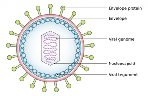
Q9. Virus Structure: Which part is similar to a cell membrane? Which part attaches to the host cell? Which part surrounds the genetic material? What kind of genetic material can a virus have?
Background
Topic: Virus Anatomy
This question tests your knowledge of virus structure and function.
Key Terms:
Envelope: Lipid layer similar to a cell membrane.
Envelope protein: Used for attachment to host cells.
Capsid: Protein shell surrounding genetic material.
Genetic material: Can be RNA or DNA.
Step-by-Step Guidance
Identify the envelope and compare it to a cell membrane.
Locate the envelope protein and describe its role in attachment.
Find the capsid and explain its function.
Note the types of genetic material viruses can have.
Try solving on your own before revealing the answer!
Final Answer:
Envelope: Similar to cell membrane; Envelope protein: Attaches to host; Capsid: Surrounds genetic material; Genetic material: RNA or DNA.
Viruses use these structures to infect and replicate.
Q10. Identify whether the symptoms described are caused by a lytic or lysogenic virus. How do you know?
Background
Topic: Viral Life Cycles
This question tests your ability to distinguish between lytic and lysogenic viral infections based on symptoms and timing.
Key Terms:
Lytic cycle: Immediate symptoms, cell lysis.
Lysogenic cycle: Dormant phase, symptoms recur over time.
Step-by-Step Guidance
Read the description and note if symptoms appear immediately or recur after a period.
Recall that lytic infections cause rapid symptoms, while lysogenic infections can remain dormant and reactivate.
Use this information to decide which cycle is responsible.
Try solving on your own before revealing the answer!
Final Answer:
Recurring symptoms indicate lysogenic; immediate symptoms indicate lytic.
The timing and recurrence help distinguish the viral cycle.
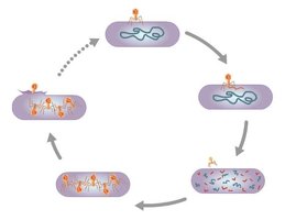
Q11. Viral Reproduction: Which cycle does this show? Describe what happens at the end of this cycle.
Background
Topic: Lytic Cycle
This question tests your understanding of the steps in viral reproduction and the outcome for the host cell.
Key Terms:
Lytic cycle: Virus replicates and causes cell to burst (lyse).
Steps: Attachment, entry, replication, assembly, release.
Step-by-Step Guidance
Identify the cycle shown (lytic) based on the steps and cell bursting.
Describe the process: virus attaches, injects genetic material, replicates, assembles new viruses, cell bursts.
Explain what happens to the host cell at the end (lysis).
Try solving on your own before revealing the answer!
Final Answer:
This shows the lytic cycle; at the end, the cell bursts and releases new viruses.
The host cell is destroyed in the process.
Q12. Which statement is correct about the cell membrane?
Background
Topic: Cell Membrane Permeability
This question tests your understanding of how the cell membrane controls movement of molecules.
Key Terms:
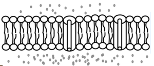
Semi-permeable: Allows some molecules to pass, not all.
Impermeable: No molecules pass.
Permeable: All molecules pass.
Step-by-Step Guidance
Recall the structure of the cell membrane (phospholipid bilayer).
Think about which molecules can pass through easily and which cannot.
Choose the statement that best describes the cell membrane's function.
Try solving on your own before revealing the answer!
Final Answer:
The cell membrane is a semi-permeable barrier, allowing some molecules to move in or out.
This is essential for cell function and homeostasis.
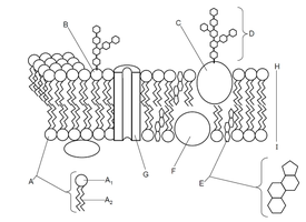
Q13. Parts of the Cell Membrane: Identify and label the components.
Background
Topic: Cell Membrane Structure
This question tests your ability to recognize and label the parts of the cell membrane.
Key Terms:
Phospholipid bilayer: Main structure of the membrane.
Proteins: Embedded in the membrane, serve various functions.
Carbohydrates: Attached to proteins/lipids, involved in cell recognition.
Cholesterol: Helps maintain membrane fluidity.
Step-by-Step Guidance
Identify the phospholipid bilayer as the main structure.
Locate embedded proteins and describe their functions.
Find carbohydrates attached to proteins/lipids.
Point out cholesterol and its role in membrane fluidity.
Try solving on your own before revealing the answer!
Final Answer:
The cell membrane includes a phospholipid bilayer, proteins, carbohydrates, and cholesterol.
Each component contributes to membrane structure and function.
