 Back
Viruses, Bacteriophages, and Prions: Structure, Function, and Biomedical Relevance
Study Guide - Smart Notes
 Tailored notes based on your materials, expanded with key definitions, examples, and context.
Non-Living Infectious Agents
Introduction to Viruses, Bacteriophages, and Prions
Non-living infectious agents such as viruses, bacteriophages (phages), and prions play significant roles in biology and medicine. Understanding their structure and mechanisms of action is essential for developing biochemical tools and therapeutic interventions.
Prions
Nature and Mechanism of Prions
Prions are infectious proteins that cause neurodegenerative diseases by inducing abnormal folding of specific normal cellular proteins, primarily in the brain. Unlike viruses and bacteria, prions contain no nucleic acids (DNA or RNA).
Definition: Prions are misfolded forms of normal proteins that can induce other normal proteins to also misfold, leading to disease.
Mechanism: The abnormal prion protein interacts with the normal protein, converting it into the prion form. This process leads to the accumulation of prion aggregates, which disrupt cell function and cause cell death.
Diseases: Prions are responsible for diseases such as Creutzfeldt-Jakob disease in humans, scrapie in sheep, and mad cow disease in cattle.
Biochemical Tools: Understanding prion propagation has informed research into protein folding and misfolding, relevant to other diseases like Alzheimer's.
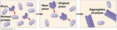
Example: In prion diseases, the accumulation of prion aggregates in neural tissue leads to spongiform changes and neurodegeneration.
Viruses
General Structure and Characteristics
Viruses are non-living infectious particles that require a host cell to replicate. They possess some characteristics of living organisms, such as the ability to evolve and reproduce, but lack independent metabolic processes.
Structure: Viruses consist of genetic material (DNA or RNA) enclosed in a protein coat called a capsid. Some viruses have an additional lipid envelope derived from the host cell membrane.
Classification: Viruses are classified based on their genetic material (DNA or RNA, single- or double-stranded) and their structure (helical, icosahedral, complex).
Host Range: Each virus has a specific host range determined by the interaction between viral surface proteins and host cell receptors.
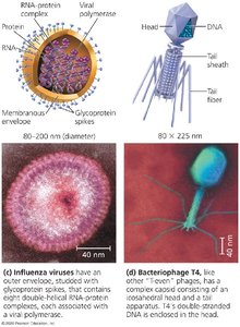
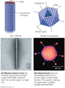
Example: The influenza virus has an outer envelope with glycoprotein spikes, while bacteriophage T4 has a complex structure with a head, tail, and tail fibers.
Viral Replication Cycles
Viruses replicate by hijacking the host cell's machinery. The general steps include attachment, entry, replication, assembly, and release.
Attachment: Virus binds to specific receptors on the host cell surface.
Entry: Viral genome enters the host cell, often by fusion or endocytosis.
Replication and Transcription: Host enzymes replicate the viral genome and transcribe viral mRNA.
Translation: Host ribosomes translate viral mRNA into viral proteins.
Assembly: New viral particles self-assemble from the replicated genome and proteins.
Release: New viruses exit the cell, often lysing the host or budding from the membrane.
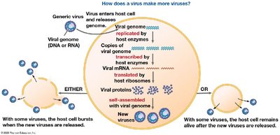
Example: Some viruses cause the host cell to burst (lyse), while others allow the host cell to survive and continue producing viruses.
Bacteriophages (Phages)
Phage Structure and Replication
Bacteriophages are viruses that infect bacteria. They have complex structures and can undergo two main types of replication cycles: lytic and lysogenic.
Lytic Cycle: The phage attaches to the bacterial cell, injects its DNA, directs the host to produce new phage components, assembles new phages, and lyses the cell to release progeny.
Lysogenic Cycle: The phage DNA integrates into the bacterial chromosome as a prophage and is replicated along with the host genome. It can later enter the lytic cycle under certain conditions.
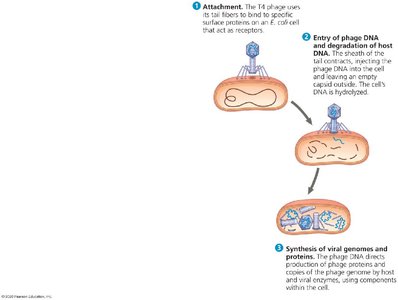
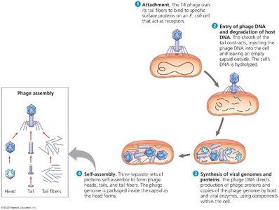
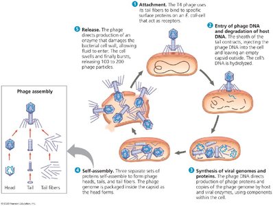
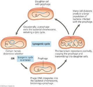
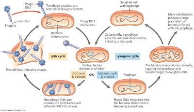
Example: Bacteriophage T4 infects Escherichia coli and can rapidly produce hundreds of new phages through the lytic cycle.
Eukaryotic Viruses
Replication in Eukaryotic Cells
Eukaryotic viruses infect animal and plant cells, often using complex entry and exit mechanisms. Their replication strategies vary depending on the type of genome (DNA or RNA).
Entry: Viruses may enter by membrane fusion or endocytosis.
Replication: DNA viruses typically replicate in the nucleus, while RNA viruses often replicate in the cytoplasm.
Assembly and Release: New virions are assembled and released by budding (enveloped viruses) or cell lysis (non-enveloped viruses).
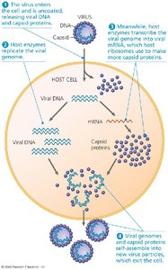
Example: The influenza virus replicates in the host cell cytoplasm and is released by budding from the plasma membrane.
Case Study: SARS-CoV-2 (Coronavirus)
Structure and Pathogenesis
Severe Acute Respiratory Syndrome Coronavirus 2 (SARS-CoV-2) is the causative agent of COVID-19. It is an enveloped, positive-sense single-stranded RNA virus with characteristic spike proteins that facilitate entry into host cells.
Infection: The virus binds to the ACE2 receptor on human cells, allowing entry and subsequent replication.
Disease: Infection can cause respiratory symptoms ranging from mild to severe, including pneumonia and acute respiratory distress syndrome.
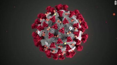
Example: The spike protein is a major target for vaccines and therapeutic antibodies.
Human Immunodeficiency Virus (HIV)
HIV Structure and Life Cycle
HIV is a retrovirus that causes Acquired Immunodeficiency Syndrome (AIDS). It targets immune cells, particularly CD4+ T cells, leading to immune system failure.
Genome: HIV contains two copies of single-stranded RNA and the enzyme reverse transcriptase.
Replication: After entry, reverse transcriptase converts viral RNA into DNA, which integrates into the host genome as a provirus.
Pathogenesis: Progressive loss of CD4+ T cells impairs immune function, increasing susceptibility to opportunistic infections.
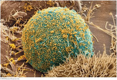
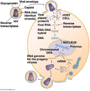
Example: Antiretroviral therapy (ART) targets multiple steps in the HIV life cycle to control infection.
Types of HIV Drugs
Several classes of drugs are used to treat HIV infection, each targeting a different stage of the viral life cycle:
Nucleoside Reverse Transcriptase Inhibitors (NRTIs): Block reverse transcription of viral RNA.
Non-nucleoside Reverse Transcriptase Inhibitors (NNRTIs): Inhibit reverse transcriptase by binding to a different site than NRTIs.
Protease Inhibitors: Prevent viral protein processing and maturation.
Integrase Inhibitors: Block integration of viral DNA into the host genome.
Fusion Inhibitors: Prevent fusion of the viral envelope with the host cell membrane.
CCR5 Antagonists: Block the CCR5 co-receptor required for HIV entry into cells.
CD4 Post-Attachment Inhibitor: Prevents post-attachment steps required for viral entry.
Example: Combination antiretroviral therapy (cART) uses drugs from multiple classes to suppress HIV replication and prevent resistance.
Summary Table: Comparison of Prions, Viruses, and Bacteriophages
Agent | Genetic Material | Structure | Replication | Diseases |
|---|---|---|---|---|
Prion | None | Misfolded protein | Induces misfolding of normal proteins | Creutzfeldt-Jakob, Mad Cow |
Virus | DNA or RNA | Capsid, sometimes envelope | Requires host cell machinery | Influenza, COVID-19, HIV/AIDS |
Bacteriophage | DNA or RNA | Complex (head, tail, fibers) | Lytic or lysogenic cycles in bacteria | Bacterial lysis |
