 Back
BackWater and Electrolyte Balance: Osmoregulation and Excretion
Study Guide - Smart Notes
 Tailored notes based on your materials, expanded with key definitions, examples, and context.
Water and Electrolyte Balance
Osmoregulation: Maintaining Internal Balance
Osmoregulation is the process by which organisms regulate the balance of water and solutes within their bodies, maintaining homeostasis despite varying environmental conditions. This process is essential for cellular function and overall survival.
Osmoregulation regulates solute concentrations and balances the gain and loss of water.
Marine organisms must regulate salt uptake and conserve water, while freshwater organisms must regulate water uptake and conserve solutes/salts.
Homeostasis ensures these balances are maintained within narrow limits.
Excretion: Removal of Metabolic Wastes
Excretion is the process of eliminating nitrogenous metabolites and other waste products resulting from the breakdown of proteins and nucleic acids. This is crucial for preventing toxic accumulation in the body.
Excretion helps maintain internal chemical balance by removing harmful substances.
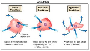
Osmosis and Osmolarity
Principles of Osmosis
Osmosis is the movement of water across a selectively permeable membrane from a region of lower solute concentration (hypoosmotic) to higher solute concentration (hyperosmotic).
Osmolarity: The total solute concentration of a solution, including both penetrating and non-penetrating solutes.
Tonicity: Refers only to non-penetrating solutes and their effect on cell volume.
Water moves from hypoosmotic to hyperosmotic solutions.
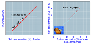
Osmoregulatory Strategies
Osmoconformers vs. Osmoregulators
Animals employ different strategies to cope with osmotic challenges in their environments.
Osmoconformers: Animals that are isoosmotic with their surroundings and do not actively regulate their internal osmolarity (e.g., most marine invertebrates).
Osmoregulators: Animals that expend energy to control water uptake and loss in hyperosmotic or hypoosmotic environments (e.g., most marine vertebrates, freshwater animals).
Stenohaline vs. Euryhaline Animals
Stenohaline: Animals that cannot tolerate substantial changes in external osmolarity (most animals).
Euryhaline: Animals that can survive large fluctuations in external osmolarity.
Osmoregulation in Aquatic Environments
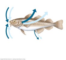
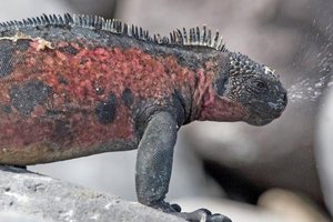
Marine Animals
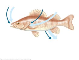
Marine vertebrates and some invertebrates are osmoregulators. Marine bony fishes are hypoosmotic to seawater, losing water by osmosis and gaining salt by diffusion and from food. They balance water loss by drinking seawater and excreting salts.
Excrete salt ions from gills and small amounts of water in urine.
Gain water and salt ions from food and drinking seawater.
Freshwater Animals
Freshwater animals constantly take in water by osmosis from their hypoosmotic environment and lose salts by diffusion. They maintain water balance by excreting large amounts of dilute urine and replace lost salts through food and uptake across the gills.
Animals in Temporary Waters: Anhydrobiosis
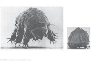
Some aquatic invertebrates can survive extreme dehydration by entering a dormant state called anhydrobiosis.
Example: Tardigrades lose almost all body water and survive until rehydration is possible.
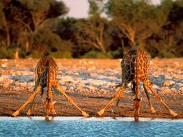
Land Animals
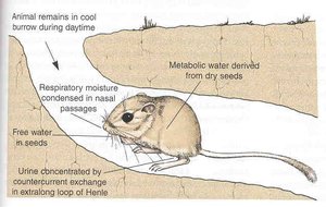
Land animals manage water budgets by drinking, eating moist foods, and using metabolic water. Desert animals have adaptations such as nocturnal lifestyles and anatomical features to minimize water loss.
Example: Kangaroo rats obtain water from seeds and produce highly concentrated urine.
Transport Epithelia in Osmoregulation
Specialized Epithelial Cells
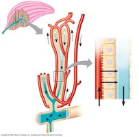
Transport epithelia are specialized cells that regulate solute movement, often arranged in complex tubular networks. An example is the salt glands of marine birds, which remove excess sodium chloride from the blood.
Nitrogenous Wastes
Ammonia
Ammonia (NH3) is highly toxic and is excreted by most aquatic organisms. It requires large amounts of water for excretion and is released across the body surface or through gills, often as ammonium (NH4+).
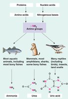
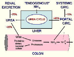
Urea
The liver of mammals and most adult amphibians converts ammonia to less toxic urea, which is transported to the kidneys for excretion. This process is energetically expensive but conserves water compared to ammonia excretion.
Uric Acid
Uric acid is excreted by birds, insects, land snails, and many reptiles. It is largely insoluble in water and is excreted as a paste, minimizing water loss. However, it is more energetically expensive to produce than urea.
Excretory Systems
Overview of Excretory Systems
Excretory systems regulate solute movement between internal fluids and the external environment. They typically involve a network of tubules and perform four main processes: filtration, reabsorption, secretion, and excretion.
Filtration: Pressure-filtering of body fluids.
Reabsorption: Reclaiming valuable solutes.
Secretion: Adding toxins and other solutes to the filtrate.
Excretion: Removing the filtrate from the system as urine.
Types of Excretory Systems
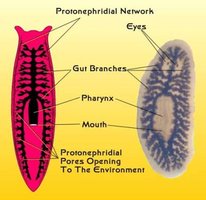
Protonephridia: Networks of dead-end tubules capped by flame bulbs, found in flatworms. They function in osmoregulation and excrete dilute fluid.
Metanephridia: Tubules that collect coelomic fluid and produce dilute urine, found in annelids like earthworms.
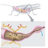
Malpighian Tubules: Remove nitrogenous wastes from hemolymph and function in osmoregulation in insects and terrestrial arthropods. No filtration occurs; instead, active transport by tubule cells produces dry waste.
Structure of the Mammalian Excretory System
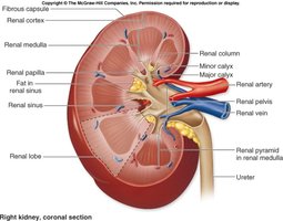
Kidney Anatomy
The mammalian excretory system consists of paired kidneys, which are the principal sites of water balance and salt regulation. Each kidney is supplied with blood by a renal artery and drained by a renal vein. Urine exits each kidney through a ureter, drains to the urinary bladder, and is expelled through the urethra.
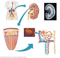
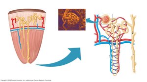
Nephron: The Functional Unit
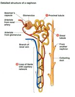
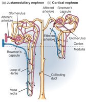
Each kidney contains about a million nephrons, each consisting of a long tubule and a ball of capillaries called the glomerulus. The nephron is responsible for filtering blood and forming urine.
Bowman's capsule surrounds the glomerulus and receives the filtrate.
There are two types of nephrons: cortical (mostly in the cortex) and juxtamedullary (extend into the medulla).
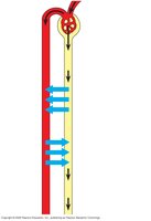
Filtration and Pathway of the Filtrate
Filtration occurs as blood pressure forces fluid from the glomerulus into Bowman's capsule. The filtrate passes through the proximal convoluted tubule (PCT), loop of Henle, and distal convoluted tubule (DCT), then into the collecting duct, which leads to the renal pelvis and ureter.
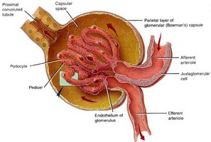

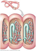
Blood Vessels Associated with Nephrons
Each nephron is supplied by an afferent arteriole, which forms the glomerular capillaries. The efferent arteriole forms peritubular capillaries around the tubules and the vasa recta around the loop of Henle, functioning as a countercurrent system.

From Blood Filtrate to Urine: Nephron Function
Proximal Convoluted Tubule (PCT)
The PCT reabsorbs ions, water, and nutrients from the filtrate into the interstitial fluid and capillaries. Some toxins are secreted into the filtrate, and the filtrate volume decreases.

Loop of Henle
Descending limb: Reabsorption of water continues via aquaporins, driven by the high osmolarity of the interstitial fluid. The filtrate becomes more concentrated.
Ascending limb: Salt diffuses out, but water cannot follow due to the absence of aquaporins. The filtrate becomes more dilute.
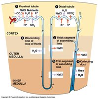

Distal Convoluted Tubule (DCT) and Collecting Duct
The DCT regulates potassium and sodium chloride concentrations, contributing to pH regulation. The collecting duct carries filtrate through the medulla to the renal pelvis, reabsorbing water, some salt, and urea, making urine hyperosmotic to body fluids.


Solute Gradients and Water Conservation
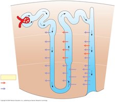
Osmotic Gradient in the Kidney
The loops of Henle and collecting ducts create an osmotic gradient in the kidney, allowing urine to be much more concentrated than blood. NaCl and urea contribute to the osmolarity of the interstitial fluid, driving water reabsorption.
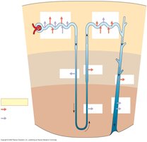
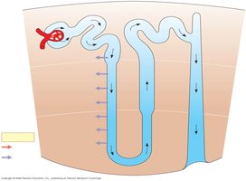

The Two-Solute Model (NaCl & Urea)
The countercurrent multiplier system in the loop of Henle maintains a high salt concentration in the kidney, allowing for the formation of concentrated urine. The vasa recta supplies nutrients without disrupting the osmolarity gradient due to countercurrent blood flow.
Adaptations in Vertebrate Kidneys
Mammals
Mammals with long loops of Henle (juxtamedullary nephrons) are better adapted for water conservation, a key adaptation for terrestrial life in dry environments.
Birds and Other Reptiles
Birds have shorter loops of Henle but conserve water by excreting uric acid as a paste. Other reptiles have only cortical nephrons and also excrete uric acid.
Regulation of Kidney Function
Antidiuretic Hormone (ADH)
ADH (vasopressin) regulates the osmolarity of urine by increasing water reabsorption in the distal tubule and collecting ducts. An increase in blood osmolarity triggers ADH release, conserving water. Alcohol inhibits ADH release, leading to increased urine output.
Renin-Angiotensin-Aldosterone System (RAAS)
The RAAS is a hormonal feedback circuit that responds to drops in blood pressure or volume. The juxtaglomerular apparatus releases renin, triggering the formation of angiotensin II, which raises blood pressure and stimulates aldosterone release, increasing salt and water reabsorption.
Atrial Natriuretic Peptide (ANP)
ANP opposes the RAAS by inhibiting renin release in response to increased blood volume and pressure, promoting excretion of salt and water to lower blood pressure.
Summary Table: Types of Nitrogenous Wastes
Waste Type | Example Organisms | Water Requirement | Relative Toxicity | Energetic Cost |
|---|---|---|---|---|
Ammonia | Most aquatic animals, bony fishes | High | Very toxic | Low |
Urea | Mammals, most amphibians, sharks | Moderate | Less toxic | Moderate |
Uric Acid | Birds, insects, reptiles, land snails | Very low | Least toxic | High |
Key Terms and Definitions
Isoosmotic: Equal osmolarity between two solutions.
Hyperosmotic: Higher solute concentration compared to another solution.
Hypoosmotic: Lower solute concentration compared to another solution.
Osmoregulator: Organism that actively regulates internal osmolarity.
Osmoconformer: Organism that matches internal osmolarity to the environment.
Stenohaline: Tolerates only narrow range of external osmolarity.
Euryhaline: Tolerates wide range of external osmolarity.
Anhydrobiosis: Dormant state allowing survival during extreme dehydration.
