 Back
Lecture 8 & 9: Protein Structure and Function
 1:39

Terms in this set (26)
Carbohydrates, lipids, proteins, and nucleic acids.
Enzymes (speed reactions)
provide structure (collagen/keratin),
transport (hemoglobin),
signaling (hormones/receptors)
defense (antibodies).
The shape dictates function; the amino acid sequence folds into a shape determining binding and activity.
Central (alpha) carbon bonded to an amino group, carboxyl group, hydrogen atom, and a side chain (R group).
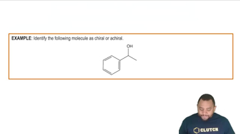
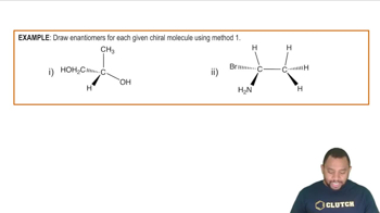
A carbon atom bonded to four different groups
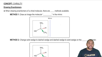
allowing for mirror-image molecules called enantiomers.
Isomers differing in bonding order of atoms
H3C-CH2-OH and H3C-O-CH3.
Isomers with same bonding order but different spatial arrangement: enantiomers (mirror images)
diastereoisomers (not mirror images, multiple chiral centers).
Nonpolar (hydrocarbons, hydrophobic)
neutral polar (contain O, N, or S, hydrophilic)
acidic (carboxylic acid, negative charge)
basic (nitrogens, positive charge).
Amino acids with both positive (amine) and negative (carboxyl) charges at pH 7, making them dipolar.
Acidic amino acids have negative charges
basic amino acids have positive charges at physiological pH.
The exact linear sequence of amino acids linked by peptide bonds,
read from N-terminus to C-terminus.
Glycine is small and flexible;
proline has a rigid ring causing kinks;
cysteine forms disulfide bonds stabilizing structure.
Alpha-helix (coiled, stabilized by H-bonds 4 residues apart) and beta-pleated sheets (strands linked by H-bonds).
Primary: peptide bonds;
Secondary: H-bonds between backbone groups;
Tertiary: hydrophobic, H-bonds, ionic, van der Waals, disulfide bonds;
Quaternary: same as tertiary but between subunits.
Collagen: fibrous, quaternary triple helix;
Keratin: fibrous, quaternary coiled bundles;
Hemoglobin: globular, quaternary with 4 subunits;
Myoglobin: globular, tertiary single chain.
Proteins with covalently bound groups: glycoproteins, lipoproteins, metalloproteins, phosphoproteins, hemoproteins, nucleoproteins.
Myoglobin stores oxygen in muscles;
hemoglobin transports oxygen in blood;
collagen provides structural support in connective tissues.
Hemoglobin molecules form long, rigid, sickle-shaped fibers causing distorted red blood cells.
Protein size (molecular mass) by denaturing proteins and giving them uniform negative charge for separation by size.
Scurvy (vitamin C deficiency) and Osteogenesis imperfecta (brittle bone disease) due to poor collagen quality or quantity.
Disulfide bonds between cysteine residues.
Proteins that assist other proteins in folding correctly within the cell.
Via autophagy (vesicle delivery to lysosomes), proteasomes (proteolysis), and lysosomes (recycling).
Prions are misfolded proteins that induce misfolding in normal proteins, causing fatal neurodegenerative diseases called transmissible spongiform encephalopathies (TSEs).
Loss of normal 3D structure due to disruption of H-bonds, ionic, and hydrophobic interactions by heat, pH changes, detergents, mechanical agitation, organic solvents, or salts.
Peptide bonds are broken by adding water, splitting the protein into smaller peptides or amino acids.