 Back
BackAmino Acids, Proteins, and Enzymes: Structure, Function, and Mechanisms
Study Guide - Smart Notes
 Tailored notes based on your materials, expanded with key definitions, examples, and context.
Amino Acids, Proteins, and Enzymes
Functions of Proteins
Proteins are essential biomolecules that perform a wide variety of functions in living organisms. They are classified based on their roles in the body:
Structural proteins: Provide support and shape to cells and tissues (e.g., collagen in tendons, keratin in hair).
Contractile proteins: Enable movement (e.g., myosin and actin in muscles).
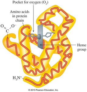
Transport proteins: Carry substances throughout the body (e.g., hemoglobin transports oxygen).
Storage proteins: Store nutrients (e.g., casein in milk, ferritin stores iron).
Hormonal proteins: Regulate physiological processes (e.g., insulin regulates blood glucose).
Enzymatic proteins: Catalyze biochemical reactions (e.g., sucrase, trypsin).
Protective proteins: Defend against foreign substances (e.g., immunoglobulins).
Amino Acids: Structure and Classification
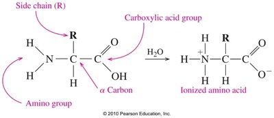
General Structure of Amino Acids
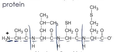
Amino acids are the building blocks of proteins. Each amino acid contains:
An amino group (–NH2)
A carboxylic acid group (–COOH)
A hydrogen atom
A unique side chain (R group) attached to the central (α) carbon
Examples of Amino Acids
Each amino acid differs by its side chain (R group). For example:
Glycine (Gly): R = H
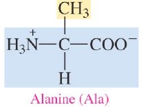
Alanine (Ala): R = CH3

Classification of Amino Acids
Amino acids are classified based on the properties of their side chains:
Nonpolar (hydrophobic): Side chains are hydrocarbons or aromatic groups (e.g., alanine, valine).
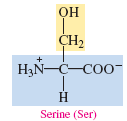
Polar (neutral, hydrophilic): Side chains contain polar groups like alcohols, thiols, or amides (e.g., serine, threonine).
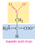
Polar (acidic): Side chains contain carboxyl groups (e.g., aspartic acid).
Polar (basic): Side chains contain amino groups (e.g., lysine).
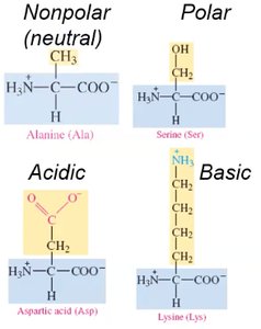
Nonpolar Amino Acids
Nonpolar amino acids have side chains that are alkyl or aromatic groups, making them hydrophobic.
Polar (Neutral) Amino Acids
Polar neutral amino acids have side chains with alcohol, thiol, or amide groups, making them hydrophilic.
Polar (Acidic) and Polar (Basic) Amino Acids
Acidic amino acids have side chains with carboxyl groups, while basic amino acids have side chains with amino groups.

Summary Table: Amino Acid Side Chains

Zwitterions and Isoelectric Points
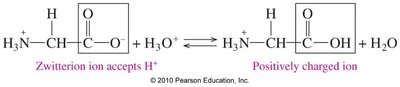
Zwitterions
In aqueous solution, amino acids exist as zwitterions, molecules with both a positive and a negative charge but overall neutral. This occurs when the amino group is protonated (–NH3+) and the carboxyl group is deprotonated (–COO–).
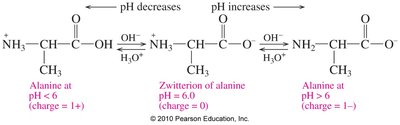
Isoelectric Point (pI)
The isoelectric point (pI) is the pH at which the amino acid exists as a zwitterion and has no net charge. For most nonpolar and polar (neutral) amino acids, the pI is between 5.1 and 6.3.
Effect of pH on Amino Acid Charge
At pH < pI: The amino acid has a net positive charge (protonated form).
At pH = pI: The amino acid is a zwitterion (net charge = 0).
At pH > pI: The amino acid has a net negative charge (deprotonated form).

Peptide Bonds and Protein Structure
Peptide Bond Formation
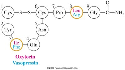
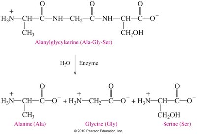
A peptide bond is an amide bond formed between the carboxyl group of one amino acid and the amino group of another. This reaction releases water and links amino acids into peptides and proteins.
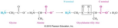

Naming Peptides
Peptides are named by giving the N-terminal amino acid a -yl ending and using the full name for the C-terminal amino acid. For example, Gly-Ala is glycylalanine.

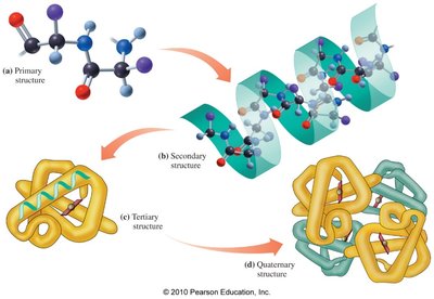
Primary Structure of Proteins
The primary structure is the unique sequence of amino acids in a protein. Even a single change in this sequence can alter protein function.
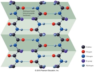
Secondary Structure
The secondary structure refers to localized folding patterns stabilized by hydrogen bonds:
Alpha helix (α-helix): A coiled structure stabilized by hydrogen bonds between every fourth amino acid.

Beta-pleated sheet (β-sheet): Polypeptide chains arranged side by side, stabilized by hydrogen bonds between chains.

Triple helix: Three polypeptide chains woven together, as in collagen.
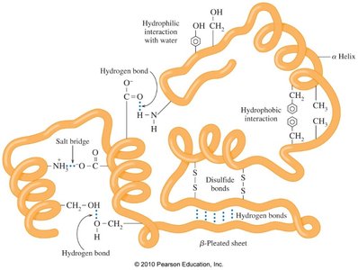
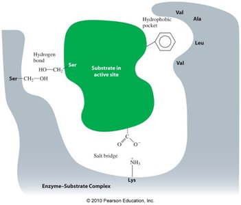
Tertiary Structure
The tertiary structure is the overall three-dimensional shape of a protein, stabilized by interactions between R groups:
Hydrophobic interactions
Hydrophilic interactions
Salt bridges (ionic bonds)
Hydrogen bonds
Disulfide bonds (covalent S–S bonds between cysteines)
Globular and Fibrous Proteins
Globular proteins: Compact, spherical, water-soluble; function in transport, metabolism, etc. (e.g., myoglobin).
Fibrous proteins: Long, fiber-like, insoluble; provide structural support (e.g., keratin, collagen).
Quaternary Structure
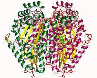
The quaternary structure is the arrangement of two or more polypeptide subunits in a functional protein (e.g., hemoglobin has four subunits).
Summary Table: Protein Structural Levels

Protein Hydrolysis and Denaturation
Protein Hydrolysis
Hydrolysis breaks peptide bonds, producing smaller peptides or amino acids. In the body, this process is catalyzed by enzymes during digestion and tissue repair.
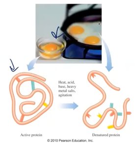
Denaturation
Denaturation disrupts the secondary, tertiary, and quaternary structures of proteins, causing loss of function. It can be caused by heat, acids, bases, heavy metals, or agitation.
Enzymes: Biological Catalysts
Enzyme Function
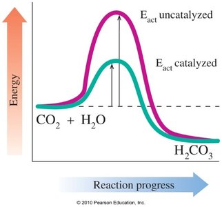
Enzymes are proteins that catalyze biochemical reactions by lowering the activation energy, thus increasing reaction rates.
Naming and Classification of Enzymes
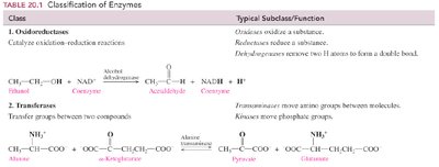
Enzyme names often end in -ase and may indicate the substrate or reaction type. Enzymes are classified by the reactions they catalyze:
Class | Type of Reaction | Example |
|---|---|---|
Oxidoreductases | Oxidation–reduction | Oxidases, dehydrogenases |
Transferases | Transfer groups | Kinases, transaminases |
Hydrolases | Hydrolysis | Peptidases, lipases |
Lyases | Add/remove atoms to/from double bonds | Decarboxylases |
Isomerases | Rearrange atoms | Isomerases, epimerases |
Ligases | Join molecules using ATP | Synthetases, carboxylases |
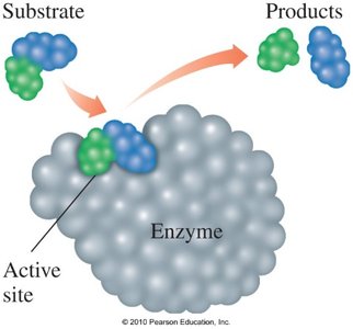
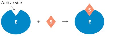
Enzyme Active Site and Mechanism
The active site is the region of the enzyme where the substrate binds and the reaction occurs. The enzyme-substrate complex forms, the reaction proceeds, and products are released.
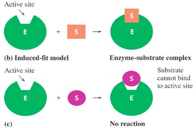
Models of Enzyme Action
Lock-and-key model: The active site is rigid; only substrates with the correct shape fit.
Induced-fit model: The active site is flexible and adjusts to fit the substrate, enhancing specificity.
Enzyme Specificity
Enzymes may be specific for a single substrate, a group of similar substrates, or a particular type of bond.
Factors Affecting Enzyme Activity
Temperature: Enzymes have an optimum temperature (usually 37°C in humans). Activity decreases at low temperatures and denaturation occurs at high temperatures.
pH: Each enzyme has an optimum pH. Activity decreases at pH values above or below the optimum due to disruption of the enzyme's structure.
Enzyme concentration: Increasing enzyme concentration increases reaction rate (if substrate is not limiting).
Substrate concentration: Increasing substrate concentration increases reaction rate until the enzyme is saturated.
Enzyme Inhibition
Competitive inhibitors: Resemble the substrate and compete for the active site. Their effect can be reversed by increasing substrate concentration.
Noncompetitive inhibitors: Bind elsewhere on the enzyme, changing its shape and preventing substrate binding. Their effect cannot be reversed by adding more substrate.
Irreversible inhibitors: Form covalent bonds with the enzyme, permanently inactivating it (e.g., cyanide, penicillin).
Diagnostic Enzymes
Elevated levels of certain enzymes in the blood can indicate tissue damage or disease (e.g., CK, LDH, and AST after a heart attack).
Summary Table: Optimum pH for Selected Enzymes
Enzyme | Location | Substrate | Optimum pH |
|---|---|---|---|
Pepsin | Stomach | Peptide Bonds | 1.5–2.0 |
Sucrase | Small Intestine | Sucrose | 6.2 |
Amylase | Pancreas | Amylose | 6.7–7.0 |
Urease | Liver | Urea | 7.0 |
Trypsin | Small Intestine | Peptide Bonds | 7.7–8.0 |
Lipase | Pancreas | Lipid (Ester Bonds) | 8.0 |
Arginase | Liver | Arginine | 9.7 |
Additional info: This guide covers the structure, classification, and function of amino acids, proteins, and enzymes, as well as the mechanisms of enzyme action and regulation, suitable for introductory college-level chemistry courses.
