 Back
Blood: Structure, Function, and Disorders – Mini-Textbook Study Notes for GOB Chemistry
Study Guide - Smart Notes
 Tailored notes based on your materials, expanded with key definitions, examples, and context.
Blood: Structure, Function, and Disorders
Functions of Blood
Blood is a vital connective tissue responsible for transport, regulation, and protection within the body. Its unique composition allows it to fulfill several essential physiological roles.
Transport: Blood delivers oxygen and nutrients to cells, removes metabolic wastes, and transports hormones.
Regulation: Maintains body temperature, pH balance (using buffers and bicarbonate ions), and fluid volume.
Protection: Prevents blood loss via clotting and defends against infection through immune agents (antibodies, complement proteins, and white blood cells).
Components of Blood
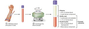
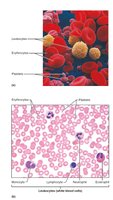
Blood consists of plasma (liquid matrix) and formed elements (cells and cell fragments). The formed elements include erythrocytes (red blood cells), leukocytes (white blood cells), and platelets.
Plasma: Straw-colored fluid containing water, proteins (albumin, globulins, fibrinogen), nutrients, gases, hormones, and waste products.
Formed Elements:
Erythrocytes: Red blood cells, responsible for gas transport.
Leukocytes: White blood cells, involved in immunity.
Platelets: Cell fragments essential for clotting.
Physical Characteristics and Volume
Blood is a viscous, opaque fluid with a metallic taste. Its color varies with oxygen content: bright red when oxygen-rich, dark red when oxygen-poor. Blood pH is tightly regulated between 7.35–7.45.
Volume: 5–6 L in males, 4–5 L in females (about 8% of body weight).
Viscosity: About five times more viscous than water, mainly due to erythrocytes.
Blood Plasma
Plasma is the fluid portion of blood, making up about 55% of its volume. It contains over 100 dissolved solutes, including nutrients, gases, hormones, wastes, proteins, and inorganic ions.
Plasma Proteins:
Albumin: Most abundant; functions as a blood buffer, carrier, and maintains osmotic pressure.
Globulins: Include antibodies and transport proteins.
Fibrinogen: Essential for blood clotting.
Formed Elements
The formed elements of blood have unique features. Erythrocytes lack nuclei and most organelles, platelets are cell fragments, and leukocytes are complete cells. Most formed elements survive only a few days and are replaced by stem cells in red bone marrow.
Erythrocytes (Red Blood Cells)
Structure and Function
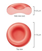
Erythrocytes are small, biconcave cells lacking a nucleus and most organelles. Their shape provides a large surface area for gas exchange and allows flexibility to pass through capillaries.
Hemoglobin: The main component, responsible for oxygen and carbon dioxide transport.
Absence of mitochondria: Prevents consumption of transported oxygen; ATP is produced anaerobically.
Hemoglobin Structure and Function
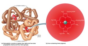
Hemoglobin is a protein composed of four globin subunits (two alpha, two beta) and four heme groups, each with a central iron ion. Each hemoglobin molecule can bind four oxygen molecules.
Oxyhemoglobin: Formed when oxygen binds to hemoglobin (bright red).
Deoxyhemoglobin: Formed when oxygen is released (dark red).
Carbaminohemoglobin: Formed when carbon dioxide binds to hemoglobin.
Erythropoiesis: Formation of Red Blood Cells
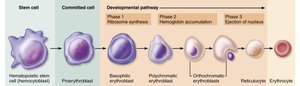
Erythropoiesis is the process of red blood cell production, occurring in red bone marrow. It involves differentiation from hematopoietic stem cells to mature erythrocytes.
Stages: Hematopoietic stem cell → myeloid stem cell → proerythroblast → basophilic erythroblast → polychromatic erythroblast → orthochromatic erythroblast → reticulocyte → erythrocyte.
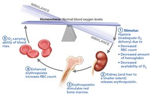
Regulation: Controlled by erythropoietin (EPO), released by kidneys in response to hypoxia.
Dietary Requirements and Fate of Erythrocytes
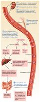
Production of erythrocytes requires amino acids, lipids, carbohydrates, vitamin B12, folic acid, and iron. Erythrocytes have a lifespan of 100–120 days; old cells are destroyed in the spleen, and their components are recycled.
Erythrocyte Disorders
Disorders include anemia (low oxygen-carrying capacity) and polycythemia (excess red blood cells).
Anemia: Caused by blood loss, insufficient production, or excessive destruction of RBCs. Types include iron-deficiency, pernicious, renal, aplastic, hemolytic, thalassemia, and sickle-cell anemia.
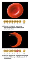
Sickle-cell anemia: Genetic disorder causing abnormal hemoglobin and crescent-shaped RBCs.
Polycythemia: Abnormal excess of RBCs, increasing blood viscosity.
Leukocytes (White Blood Cells)
Structure and Function
Leukocytes are complete cells with nuclei and organelles, crucial for defense against disease. They move through tissues by diapedesis and chemotaxis.
Leukocytosis: Increased WBC count, often in response to infection.
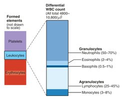
Classification: Granulocytes (neutrophils, eosinophils, basophils) and agranulocytes (lymphocytes, monocytes).
Granulocytes
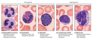
Granulocytes contain cytoplasmic granules and have lobed nuclei. They include:
Neutrophils: Most numerous, phagocytic, kill bacteria.
Eosinophils: Destroy parasitic worms, modulate immune response.
Basophils: Release histamine, involved in inflammation.
Agranulocytes
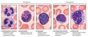
Agranulocytes lack visible granules and have spherical or kidney-shaped nuclei. They include:
Lymphocytes: Second most numerous, crucial for immunity. T cells attack infected cells; B cells produce antibodies.
Monocytes: Largest WBCs, differentiate into macrophages, active in phagocytosis.
Leukopoiesis: Formation of White Blood Cells
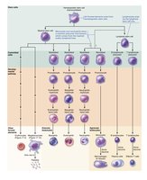
Leukopoiesis is stimulated by interleukins and colony-stimulating factors. Hematopoietic stem cells differentiate into myeloid or lymphoid stem cells, producing various WBCs.
Leukocyte Disorders
Disorders include leukopenia (low WBC count), leukemias (cancerous overproduction), and infectious mononucleosis (viral disease).
Platelets
Structure and Function
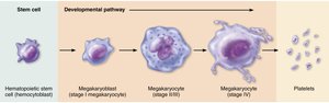
Platelets are cell fragments derived from megakaryocytes, essential for blood clotting. They form temporary plugs to seal vessel breaks.
Formation: Regulated by thrombopoietin; megakaryocytes fragment into platelets in bone marrow and lungs.
Lifespan: Platelets age quickly and degenerate in about 10 days.
Hemostasis: Prevention of Blood Loss
Steps of Hemostasis
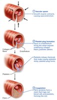
Hemostasis is a rapid, localized process to stop bleeding, involving three steps:
Vascular Spasm: Vasoconstriction reduces blood loss.
Platelet Plug Formation: Platelets adhere to exposed collagen, become activated, and aggregate.
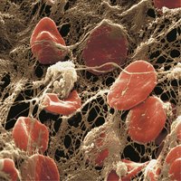
Coagulation: Reinforces plug with fibrin mesh, involving a cascade of clotting factors.
Coagulation Pathways
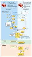
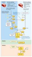
Coagulation occurs via intrinsic and extrinsic pathways, both leading to the formation of prothrombin activator, which converts prothrombin to thrombin. Thrombin then converts fibrinogen to fibrin, forming the clot.
Intrinsic Pathway: Triggered by factors within blood.
Extrinsic Pathway: Triggered by tissue factor outside blood.

Clot Retraction and Fibrinolysis
After clot formation, actin and myosin in platelets contract to stabilize the clot. Fibrinolysis removes the clot after repair, with plasmin digesting fibrin.
Disorders of Hemostasis
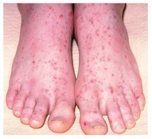
Hemostatic disorders include thromboembolic conditions (undesirable clot formation) and bleeding disorders (impaired clot formation).
Thrombocytopenia: Low platelet count, causing spontaneous bleeding.
Hemophilia: Hereditary deficiency of clotting factors.
Disseminated Intravascular Coagulation (DIC): Both widespread clotting and severe bleeding.
Blood Transfusion and Blood Groups
Blood Groups and Typing
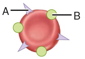
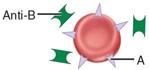
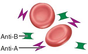
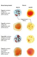
Blood groups are determined by antigens on RBC membranes. The ABO and Rh groups are most clinically significant.
ABO Groups:
Type A: A antigen, anti-B antibody
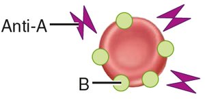
Type B: B antigen, anti-A antibody
Type AB: A and B antigens, no antibodies
Type O: No antigens, both anti-A and anti-B antibodies
Rh Group: Presence or absence of D antigen; Rh+ or Rh-
Transfusion Reactions
Transfusion of incompatible blood can cause agglutination and destruction of donor RBCs, leading to potentially fatal reactions. Group O is considered the universal donor, and group AB the universal recipient, but other antigens may cause reactions.
Blood Tests and Developmental Aspects
Blood Tests
Blood tests provide insights into health, including hematocrit, blood glucose, differential WBC count, prothrombin time, and complete blood count (CBC).
Developmental Aspects
Fetal blood cells form in yolk sac, liver, and spleen; red bone marrow becomes primary site by the seventh month. Hemoglobin F in fetuses has higher oxygen affinity than adult hemoglobin A.
Summary Table: Blood Cell Types
Cell Type | Main Function | Key Features |
|---|---|---|
Erythrocyte | O2 and CO2 transport | Biconcave, anucleate, hemoglobin-rich |
Leukocyte | Immunity | Nucleated, various types (granulocytes, agranulocytes) |
Platelet | Clotting | Cell fragments, contain clotting chemicals |
Key Equations
Hemoglobin Oxygen Binding:
Conversion of Prothrombin to Thrombin:
Conversion of Fibrinogen to Fibrin:
Additional info: These notes expand on the original lecture slides with academic context, definitions, and examples to ensure completeness and clarity for GOB Chemistry students.
