 Back
Carbohydrates and Stereochemistry: Structure, Function, and Isomerism
Study Guide - Smart Notes
 Tailored notes based on your materials, expanded with key definitions, examples, and context.
Carbohydrates and Stereochemistry
Introduction to Stereochemistry
Stereochemistry is the study of the three-dimensional properties and shapes of molecules, which is crucial for understanding the behavior and function of carbohydrates in biological systems. Stereoisomers are molecules that differ only in the spatial arrangement of their atoms, often due to the presence of chiral centers.
Stereoisomers: Molecules with the same molecular formula but different 3D arrangements.
Chiral Center: Typically a carbon atom bonded to four different groups.
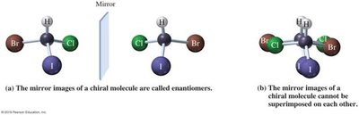
Chirality: A chiral object or molecule cannot be superimposed on its mirror image.
Achiral: Objects or molecules with a plane of symmetry; their mirror image is superimposable.
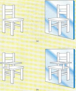
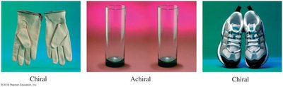
Chirality in Molecules
Chirality in molecules arises when a carbon atom is attached to four different groups, creating a stereogenic center. The presence or absence of symmetry determines whether a molecule is chiral or achiral.
Enantiomers: Non-superimposable mirror images of chiral molecules.
Physical Properties: Enantiomers share identical non-chiral properties (melting point, boiling point) but differ in chiral properties (optical activity, interactions with chiral molecules).
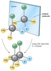
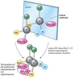
Biological Importance of Stereochemistry
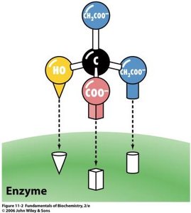
Biological molecules are stereospecific, meaning their function depends on their stereochemistry. Enzymes and receptors often distinguish between enantiomers, leading to different physiological effects.
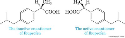
Example: The enantiomers of ibuprofen have different biological activities; only one is pharmacologically active.
Optical Activity and Nomenclature
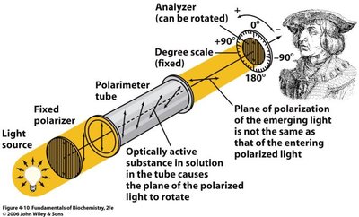
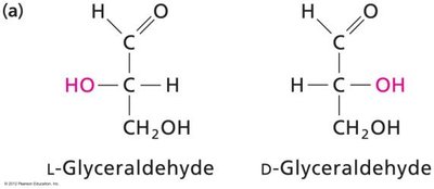
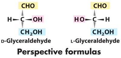
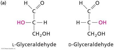
Chiral molecules are optically active, meaning they rotate plane-polarized light. The direction of rotation is denoted as levorotatory (L, left, -) or dextrorotatory (D, right, +). The L/D nomenclature is based on the structure of L-glyceraldehyde.
Polarimetry: Used to measure optical rotation.
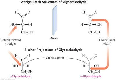
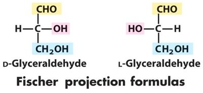
Glyceraldehyde: Reference molecule for D/L assignment.
Fischer Projections and Stereocenters
Fischer projections are a two-dimensional representation of three-dimensional molecules, especially useful for sugars. The number of possible stereoisomers for a molecule with n stereocenters is given by .
Fischer Projection: Horizontal lines represent bonds projecting above the plane; vertical lines project behind.
Stereoisomers: For n stereocenters, possible isomers.
Enantiomers: Mirror images.
Diastereomers: Stereoisomers that are not mirror images.
Carbohydrates: Classification and Structure
Carbohydrates are organic molecules with the general formula (C•H2O)n. They are classified based on the number of monomer units and the type of carbonyl group present.
Monosaccharide: Single sugar unit.
Oligosaccharide: Short chain of monosaccharides.
Polysaccharide: Long chain of monosaccharides.
Glycoprotein: Oligosaccharide linked to protein.
Glycolipid: Oligosaccharide linked to lipid.
Types of Sugars: Aldoses and Ketoses
Sugars are classified as aldoses or ketoses based on the location of the carbonyl group.
Aldose: Carbonyl group at the end (aldehyde).
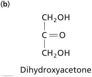
Ketose: Carbonyl group in the middle (ketone).
Triose, Tetrose, Pentose, Hexose: Number of carbon atoms (3, 4, 5, 6).
Stereochemistry of Sugars
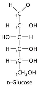
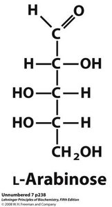
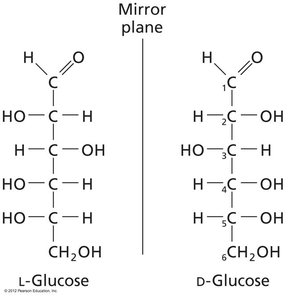
The D/L configuration of sugars is determined by the chiral center furthest from the carbonyl group, compared to glyceraldehyde. D-sugars are more common in nature.
Example: D-glucose and L-glucose are enantiomers.
Epimers and Isomerism
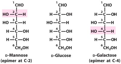
Epimers are sugars that differ only in the configuration around one specific carbon atom. Diastereomers are stereoisomers that are not mirror images.
Example: D-mannose and D-glucose are epimers at C-2; D-galactose and D-glucose are epimers at C-4.
Ring Structures: Furanose and Pyranose
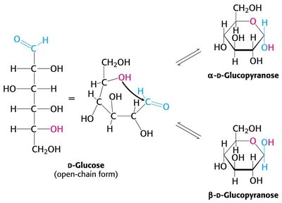
Under physiological conditions, pentoses and hexoses form ring structures by intramolecular reactions. Furanose rings have five members, while pyranose rings have six.
Furanose: Five-membered ring.
Pyranose: Six-membered ring.
Formation of Hemiacetals and Hemiketals
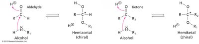
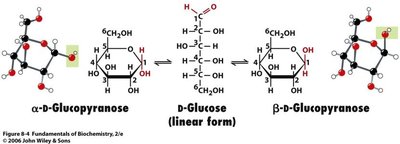
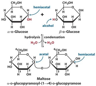
Sugar molecules convert from their linear form to ring structures by nucleophilic attack of the carbonyl by an alcohol, forming a hemiacetal (aldose) or hemiketal (ketose). The carbonyl carbon becomes the anomeric carbon, a new stereocenter.
Anomeric Carbon: The carbonyl carbon in the linear form, becomes a new chiral center in the ring.
Haworth Projections and Anomers
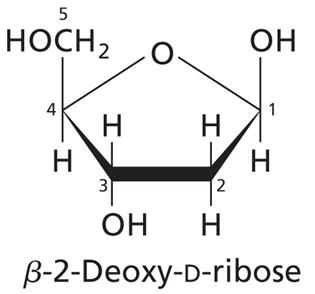
Haworth projections are simplified drawings of ring structures. Anomers are stereoisomers that differ only at the anomeric carbon. The α-anomer has the OH group on the opposite face from the CH2OH group; the β-anomer has them on the same face.
Mutarotation: Interconversion between α and β anomers via the linear form.
Reducing and Nonreducing Sugars
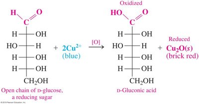
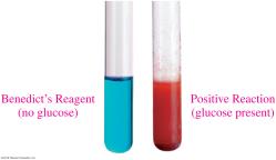
Reducing sugars have a free anomeric carbon that can act as a reducing agent. Nonreducing sugars have their anomeric carbon involved in a glycosidic bond and cannot be oxidized.
Example: Glucose is a reducing sugar; sucrose is nonreducing.
Monosaccharide Derivatives
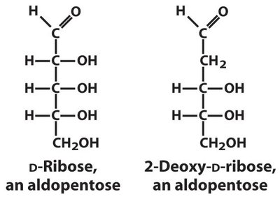
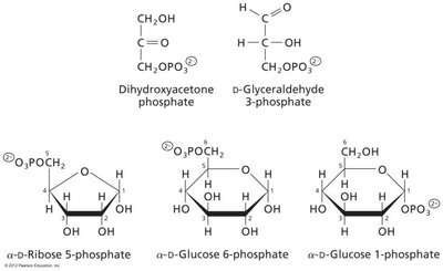
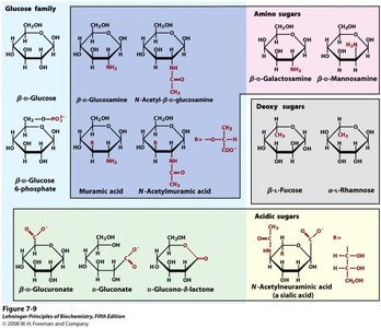
Monosaccharides can be modified by removal of hydroxyl groups (deoxy sugars) or addition of phosphate groups (phosphorylated sugars). These derivatives are important in metabolism and nucleic acid structure.
Example: 2-deoxy-D-ribose is a component of DNA.
Phosphorylated sugars: Glucose 6-phosphate, ribose 5-phosphate.
Glycosidic Bonds
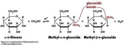
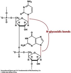
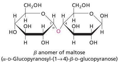
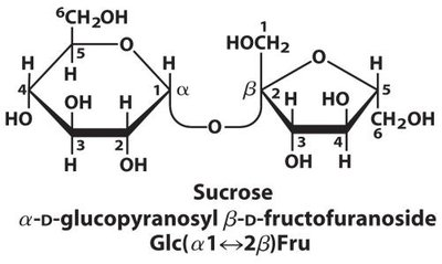
Glycosidic bonds are formed when the anomeric group of a sugar reacts with another alcohol or amine. Once formed, the anomers can no longer interconvert. Glycosidases are enzymes that cleave glycosidic bonds.
N-glycosidic bonds: Formed when sugars are linked through nitrogen, important in nucleic acids.
Disaccharides and Polysaccharides
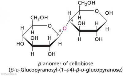
Disaccharides are formed by linking two monosaccharides via a glycosidic bond. Polysaccharides are long chains of monosaccharide units, which may be linear or branched.
Maltose: α(1→4) linkage of glucose units.
Cellobiose: β(1→4) linkage of glucose units.
Sucrose: α-D-glucopyranosyl β-D-fructofuranoside (nonreducing).
Lactose: β(1→4) linkage of galactose and glucose.
Polysaccharides: Structure and Function
Polysaccharides are classified as homopolysaccharides (same monosaccharide unit) or heteropolysaccharides (different units). Their structure determines their function, such as energy storage or structural support.
Cellulose: β(1→4) linked glucose; structural glycan in plants.
Chitin: β(1→4) linked N-acetylglucosamine; structural glycan in fungi and arthropods.
Starch (Amylose): α(1→4) linked glucose; energy storage in plants.
Amylopectin: Branched starch with α(1→6) linkages.
Glycogen: Highly branched glucose polymer in animals.
Polysaccharide | Monomer | Linkage | Function |
|---|---|---|---|
Cellulose | Glucose | β(1→4) | Structural (plants) |
Chitin | N-acetylglucosamine | β(1→4) | Structural (fungi, arthropods) |
Amylose | Glucose | α(1→4) | Energy storage (plants) |
Amylopectin | Glucose | α(1→4), α(1→6) | Energy storage (plants, branched) |
Glycogen | Glucose | α(1→4), α(1→6) | Energy storage (animals, highly branched) |
Summary Table: Key Carbohydrate Structures
Monosaccharide | Type | Structure |
|---|---|---|
Glyceraldehyde | Aldose (triose) | CH2OH-CHOH-CHO |
Dihydroxyacetone | Ketose (triose) | CH2OH-CO-CH2OH |
Ribose | Aldose (pentose) | CH2OH-(CHOH)3-CHO |
Glucose | Aldose (hexose) | CH2OH-(CHOH)4-CHO |
Mannose | Aldose (hexose) | Epimer of glucose at C-2 |
Galactose | Aldose (hexose) | Epimer of glucose at C-4 |
Fructose | Ketose (hexose) | CH2OH-CO-(CHOH)3-CH2OH |
Additional info: Students should be able to recognize and draw Fischer and Haworth projections for these sugars, identify enantiomers, diastereomers, and epimers, and understand the biological significance of carbohydrate stereochemistry.
