Back
BackChapter 10: Nuclear Chemistry – Study Notes
Study Guide - Smart Notes
 Tailored notes based on your materials, expanded with key definitions, examples, and context.
Chapter 10: Nuclear Chemistry
10.1 Introduction to Nuclear Chemistry
Nuclear chemistry focuses on the structure and behavior of atomic nuclei, including the processes by which unstable nuclei change and emit radiation. Understanding atomic number, mass number, and isotopes is foundational to this field.
Atomic Number (Z): The number of protons in the nucleus of an atom.
Mass Number (A): The total number of protons and neutrons in the nucleus.
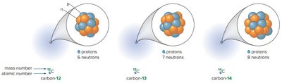
Isotopes: Atoms of the same element with different numbers of neutrons.
Radioisotopes: Unstable isotopes that spontaneously emit energy to form a more stable nucleus.
Radioactivity: The emission of nuclear radiation by a radioactive isotope.
10.1B Types of Radiation
Radioactive decay can emit several types of radiation, each with distinct properties and effects.
Alpha (α) Particles: High-energy particles containing 2 protons and 2 neutrons; charge +2, mass number 4. Symbol: or .
Beta (β) Particles: High-energy electrons; charge -1, negligible mass. Symbol: or .
Positrons: Antiparticles of beta particles; charge +1, negligible mass. Symbol: or .
Gamma (γ) Rays: High-energy electromagnetic radiation; no mass, no charge. Symbol: .
Type of Radiation | Symbol | Charge | Mass Number |
|---|---|---|---|
Alpha particle | or | +2 | 4 |
Beta particle | or | -1 | 0 |
Positron | or | +1 | 0 |
Gamma ray | 0 | 0 |
10.2 Nuclear Reactions
Nuclear reactions involve changes in the nucleus and are described by nuclear equations. The sum of mass numbers and atomic numbers must be equal on both sides of the equation.
Radioactive Decay: The process by which an unstable nucleus emits radiation to become more stable.
Nuclear Equation:
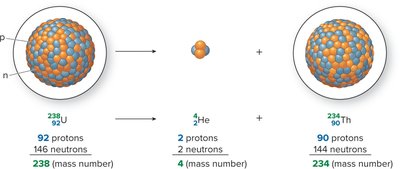
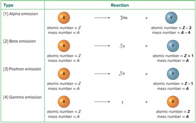
10.2A Alpha Emission
Alpha emission is the decay of a nucleus by emitting an alpha particle. The atomic number decreases by 2 and the mass number by 4.
Example:
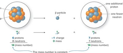
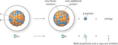
10.2B Beta Emission
Beta emission occurs when a neutron is converted to a proton and a beta particle is emitted. The atomic number increases by 1, mass number remains unchanged.
Example:
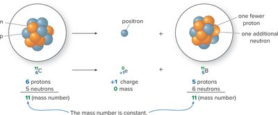
10.2C Positron Emission
Positron emission occurs when a proton is converted to a neutron and a positron is emitted. The atomic number decreases by 1, mass number remains unchanged.
Example:
10.2D Gamma Emission
Gamma emission involves the release of energy from a nucleus in the form of gamma rays. There is no change in atomic or mass number.
Example:
Gamma emission often accompanies alpha or beta emission.
Summary Table: Types of Nuclear Reactions
Type | Reaction |
|---|---|
Alpha emission | |
Beta emission | |
Positron emission | |
Gamma emission |
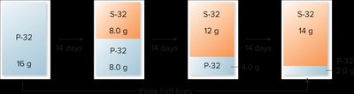
10.3 Half-Life
The half-life () of a radioactive isotope is the time required for half of a sample to decay. It is a constant property for each isotope and is independent of sample amount, temperature, and pressure.
Calculation: After each half-life, half of the remaining radioactive atoms decay.
Formula: where is the number of half-lives elapsed.
Example: If the half-life of iodine-131 is 8.0 days, after 32 days (4 half-lives), remains.
Applications of Half-Life
Radiocarbon Dating: Uses the half-life of carbon-14 to determine the age of carbon-containing materials. Living organisms maintain a constant ratio of C-14 to C-12; after death, C-14 decays and the ratio decreases, allowing age determination.
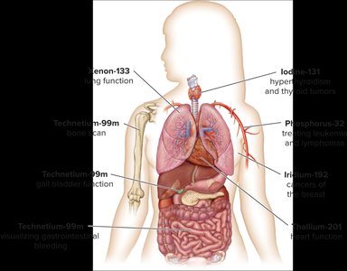
Medical and Industrial Uses: Different radioisotopes are used for dating, therapy, and imaging.
Radioisotope | Symbol | Half-Life | Use |
|---|---|---|---|
Carbon-14 | years | Archaeological dating | |
Cobalt-60 | 5.27 years | Cancer therapy | |
Iodine-131 | 8.02 days | Thyroid therapy | |
Potassium-40 | years | Geological dating | |
Phosphorus-32 | 14.3 days | Leukemia treatment | |
Technetium-99m | 6.01 hours | Organ imaging | |
Uranium-235 | years | Nuclear reactors |
10.4 Detecting and Measuring Radioactivity
Radioactivity is measured by the number of decays per second. Common units include the Curie (Ci) and the becquerel (Bq).
1 Curie (Ci): disintegrations per second
1 becquerel (Bq): 1 disintegration per second
Rad (radiation absorbed dose): Amount of radiation absorbed by 1 gram of substance
Rem (radiation equivalent for man): Accounts for energy and tissue damage potential
Radiation can damage or kill rapidly dividing cells, making it useful for cancer treatment but also hazardous. Food irradiation uses gamma rays to kill organisms in food, extending shelf life without making the food radioactive.
Biological Effects: Doses below 25 rem have no detectable effects; 25–100 rem causes temporary white blood cell decrease; >100 rem causes radiation sickness; 500 rem is lethal to 50% of humans (LD50).
10.5 Medical Applications of Radioisotopes
Diagnosis
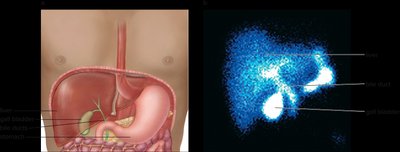
Radioisotopes are used to diagnose organ function and detect tumors. For example, technetium-99m is used for gall bladder imaging, and thallium-201 for cardiac stress tests.
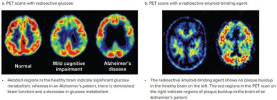
Positron Emission Tomography (PET)
PET scans use positron-emitting radioisotopes to visualize organ function, detect tumors, and monitor diseases such as Alzheimer's and cancer.
10.6 Nuclear Fission and Fusion
Nuclear Fission
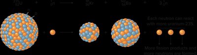
Nuclear fission is the splitting of a heavy nucleus (e.g., uranium-235) into lighter nuclei, releasing energy and neutrons. The released neutrons can initiate further fission, creating a chain reaction. Fission is used in nuclear power plants to generate electricity.
Critical Mass: The minimum amount of fissionable material needed to sustain a chain reaction.
Equation Example:
Nuclear Fusion
Nuclear fusion is the joining of two light nuclei (e.g., deuterium and tritium) to form a heavier nucleus, releasing even more energy than fission. Fusion powers the sun but requires extremely high temperatures and pressures, making it currently impractical for energy production on Earth.
Equation Example:
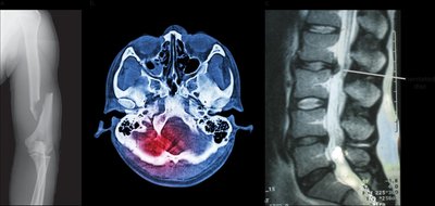
10.7 Medical Imaging without Radioactivity
Several imaging techniques do not use radioactivity:
X-rays: Electromagnetic radiation used to image bones and internal organs.
CT (Computed Tomography) Scans: Use X-rays to create cross-sectional images of the body.
MRI (Magnetic Resonance Imaging): Uses radio waves and magnetic fields to image soft tissues with minimal cellular damage.