 Back
Nucleic Acid Structure: DNA, RNA, and Their Biological Functions
Study Guide - Smart Notes
 Tailored notes based on your materials, expanded with key definitions, examples, and context.
Nucleotides: The Building Blocks of Nucleic Acids
Structural Components of Nucleotides
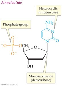
Nucleotides are the fundamental units of nucleic acids, consisting of three main components: a heterocyclic nitrogenous base, a five-carbon sugar (ribose or deoxyribose), and a phosphate group. These components are joined together through condensation reactions.
Heterocyclic nitrogenous base: Aromatic, planar, and capable of hydrogen bonding. Two types: purines (adenine, guanine) and pyrimidines (cytosine, thymine, uracil).
Sugar: Ribose (in RNA) or deoxyribose (in DNA).
Phosphate group: Provides negative charge and links nucleotides via phosphodiester bonds.
Example: The nucleotide adenosine monophosphate (AMP) contains adenine, ribose, and a single phosphate group.
Nucleosides vs. Nucleotides
A nucleoside consists of a nitrogenous base and a sugar, while a nucleotide also includes one or more phosphate groups.
Nucleoside: Base + sugar
Nucleotide: Base + sugar + phosphate
Nucleic Acids: DNA and RNA
Polynucleotide Formation
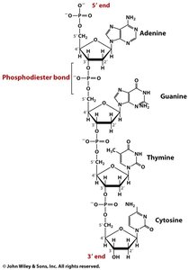
Nucleic acids are linear polymers of nucleotides, connected by phosphodiester bonds between the 3' hydroxyl of one sugar and the 5' phosphate of the next. The sequence is written from the 5' to 3' direction.
Phosphodiester backbone: Provides structural stability and directionality.
Sequence notation: 5' AGTC 3'
DNA vs. RNA: Key Differences
DNA and RNA are the two main types of nucleic acids, differing in their sugar, bases, and structure.
Feature | DNA | RNA |
|---|---|---|
Sugar | Deoxyribose | Ribose |
Fourth Base | Thymine (T) | Uracil (U) |
Strandedness | Double-stranded | Single-stranded |
3D Structure of DNA: The Double Helix
Key Structural Features
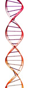
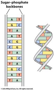
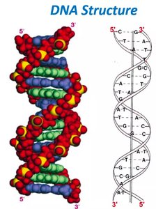
Double-stranded DNA forms a double helix, with two antiparallel strands. The phosphate backbone is on the outside, and the bases are stacked perpendicular to the backbone, forming the steps of a spiral staircase.
Antiparallel strands: One strand runs 5' to 3', the other 3' to 5'.
Phosphate backbone: Negatively charged, faces outward.
Base stacking: Planar bases provide stability via dispersion forces.
Base Pairing and Stability
Bases pair across the helix via hydrogen bonds, providing specificity and stability. Adenine pairs with thymine (2 H-bonds), and guanine pairs with cytosine (3 H-bonds).
Base Pair | Number of H-bonds |
|---|---|
A-T | 2 |
G-C | 3 |
Protein-DNA Interactions
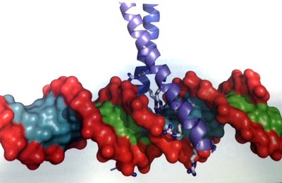
Proteins interact with DNA primarily through ionic interactions between positively-charged amino acids (arginine, lysine, histidine) and the negatively-charged phosphate backbone. Sequence-specific recognition occurs via unique patterns of hydrogen bond donors and acceptors in the major groove.
Salt bridges: Formed between basic amino acids and DNA phosphates.
Major groove: Site of sequence-specific protein binding.
DNA Organization in the Cell
Genes, Chromatin, and Chromosomes
DNA is compacted in the cell nucleus as chromatin, which consists of DNA wound around histone proteins to form nucleosomes. Chromatin is further organized into chromosomes during cell division. Genes are segments of DNA that encode proteins.
Chromatin: Orderly tangle of DNA and protein.
Nucleosome: DNA wrapped around histone proteins.
Chromosome: Highly compacted DNA-protein complex.
Complementarity and Base-Pairing
Chargaff’s Rules and DNA Complementarity
Chargaff’s rules state that in double-stranded DNA, the number of adenine bases equals thymine, and the number of guanine equals cytosine. This complementarity allows for accurate replication and information transfer.
Reverse complement: The sequence of one strand determines the sequence of the other, written in the opposite direction.
Replication: Each strand serves as a template for a new strand.
Mutations in DNA
Causes and Consequences of Mutations
A mutation is a change in the DNA sequence, often caused by mutagens such as radiation or chemicals. Mutations can alter base-pairing and, if not repaired, are propagated during DNA replication. Cytosine deamination is a common mutation, leading to uracil formation, which is recognized and repaired by cellular enzymes.
Mutagen: Substance causing DNA mutation.
Semiconservative replication: Each new DNA molecule contains one old and one new strand.
Central Dogma: DNA is replicated, transcribed to RNA, and translated to protein.
Summary Table: DNA vs. RNA vs. Nucleotides
Component | DNA | RNA | Nucleotide |
|---|---|---|---|
Sugar | Deoxyribose | Ribose | Ribose/Deoxyribose |
Bases | A, G, C, T | A, G, C, U | A, G, C, T/U |
Phosphate | Yes | Yes | Yes |
Structure | Double-stranded | Single-stranded | Monomer |
Key Equations and Concepts
Phosphodiester bond formation:
Base-pairing:
Central Dogma:
Additional info: These notes expand on the lecture slides and images, providing definitions, examples, and structural context for nucleic acids, their organization, and their biological functions. The included images directly reinforce the structural and functional explanations of nucleotides, DNA, and protein-DNA interactions.
