 Back
Protein Structure and Function: GOB Chemistry Study Notes
Study Guide - Smart Notes
 Tailored notes based on your materials, expanded with key definitions, examples, and context.
Protein Structure and Function
Levels of Protein Structure
Proteins are complex biomolecules whose function is determined by their structure. The structure of proteins is organized into four hierarchical levels:
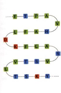
Primary Structure: The sequence of amino acids in a polypeptide chain, held together by peptide bonds.
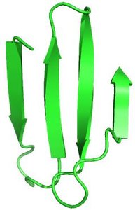
Secondary Structure: Local folding patterns such as α-helices and β-sheets, stabilized by hydrogen bonds.
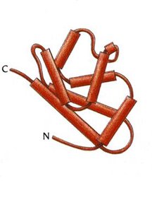
Tertiary Structure: The overall three-dimensional shape of a single polypeptide chain, stabilized by interactions among side chains.
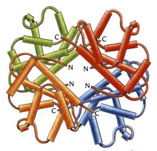
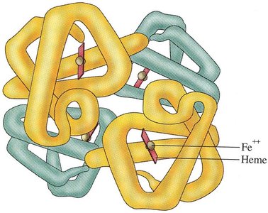
Quaternary Structure: The arrangement of multiple polypeptide chains (subunits) into a functional protein complex.
Shape Determining Interactions
The folding and stability of proteins are determined by several types of interactions:
Covalent Bonds: Peptide bonds and disulfide bonds (S-S) between cysteine residues.
Electrostatic Interactions: Ionic bonds (salt bridges) between charged side chains.
Hydrogen Bonds: Occur between hydrogen donors (O-H, N-H) and acceptors (O, N).
Van der Waals Forces: Weak interactions between all atoms.
Classification of Protein Structures
Proteins are classified based on their shape and solubility:
Globular Proteins: Compact, globe-like, typically water-soluble due to hydrophilic residues on the surface. Examples: enzymes, myoglobin, hemoglobin.
Fibrous Proteins: Elongated, insoluble, with tightly packed hydrophobic residues. Examples: collagen, keratin (hair, teeth, fingernails).
Conjugated Proteins
Conjugated proteins require non-protein components for function. The main types are:
Glycoproteins: Covalently bound carbohydrates (e.g., cell surface receptors).
Lipoproteins: Covalently bound lipids (e.g., cholesterol transporters).
Metalloproteins: Contain metal ions for structural or catalytic roles (e.g., zinc-stabilized proteins).
Phosphoproteins: Covalently bound phosphate groups for regulation or binding.
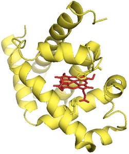
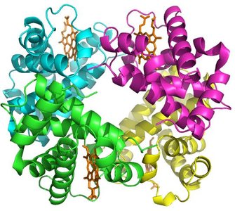
Hemoproteins: Contain heme groups for oxygen transport or catalysis.
Nucleoproteins: Contain nucleic acids (e.g., ribosomes).
Myoglobin and Hemoglobin
Structure and Function
Myoglobin and hemoglobin are globular conjugated proteins involved in oxygen transport:
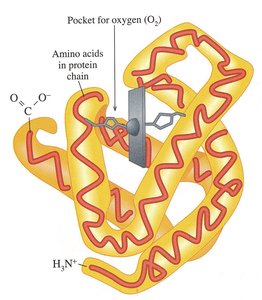
Myoglobin: Found in muscles, consists of a single polypeptide chain (tertiary structure), binds oxygen for storage.
Hemoglobin: Found in red blood cells, consists of four polypeptide chains (quaternary structure), transports oxygen from lungs to tissues.
Oxygen Binding and Transfer
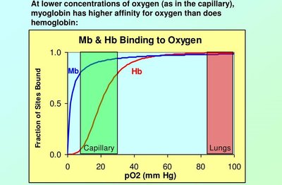
Hemoglobin undergoes conformational changes upon oxygen binding, enabling cooperative oxygen transport. Myoglobin has a higher affinity for oxygen at low concentrations, facilitating oxygen transfer from hemoglobin to muscle tissues.
Sickle Cell Anemia
Structural Changes and Disease
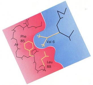
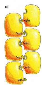
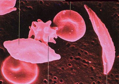
Sickle cell anemia is caused by a mutation in hemoglobin where glutamic acid (Glu) is replaced by valine (Val) at position 6. This change introduces a hydrophobic residue, leading to aggregation of hemoglobin molecules and distortion of red blood cells.
Symptoms: Extreme pain, fatigue, stroke, and anemia.
Diagnosis: Blood tests, amniocentesis.
Treatment: Hydration, antibiotics, blood transfusions, gene therapy, bone marrow transplant.
Electrophoresis: Analytical Tool
Principle and Application
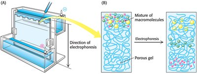
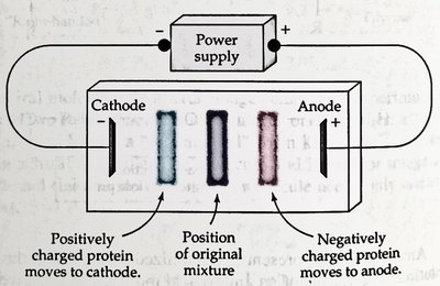
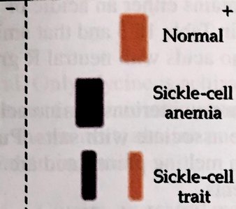
Electrophoresis is a technique used to separate proteins based on their charge and molecular weight. It is useful for diagnosing diseases such as sickle cell anemia by comparing migration patterns of normal and mutant hemoglobin.
Proteins migrate in an electric field: Negatively charged proteins move toward the anode, positively charged toward the cathode.
Diagnostic use: Sickle cell hemoglobin migrates differently due to its altered charge.
Collagen: Structure and Diseases
Structure and Function
Collagen is a fibrous protein with a quaternary structure, found in connective tissues, bones, and teeth. Its structure is stabilized by hydrogen bonds and covalent disulfide bonds. Every third residue is glycine, allowing tight packing of strands.
Subunit: Tropocollagen.
Stabilization: Hydrogen bonds and disulfide bonds.
Diseases: Osteogenesis imperfecta (brittle bone disease), scurvy (vitamin C deficiency).
Keratin: Structure and Stabilization
α-Keratin and β-Keratin
Keratin is a fibrous protein found in hair, nails, and skin. α-Keratin is stabilized by disulfide bonds, while β-Keratin is stabilized by van der Waals interactions.
α-Keratin: Disulfide bonds provide strength and stability.
β-Keratin: Van der Waals interactions provide springiness.
Protein Folding and Chaperones
Chaperones
Chaperones are proteins and protein complexes that assist in the folding of other proteins, preventing aggregation and ensuring proper function.
Prions: Misfolded Infectious Proteins
Prion Diseases
Prions are misfolded proteins in the central nervous system that induce other proteins to misfold, causing transmissible spongiform encephalopathies (TSEs). These diseases are rapidly progressive and fatal, affecting both humans and animals.
Protein Degradation and Denaturation
Protein Degradation
Proteins are degraded by breaking peptide bonds through processes such as autophagy, lysosomal degradation, and proteasome activity. Protein hydrolysis breaks peptide bonds, releasing amino acids.
Protein Denaturation
Denaturation is the unfolding of proteins, caused by heat, mechanical agitation, detergents, organic solvents, pH changes, and inorganic salts. Denaturation disrupts non-covalent interactions, leading to loss of function.
Agents of denaturation: Heat, mechanical agitation, detergents, organic compounds, pH change, inorganic salts.
Summary Table: Protein Structure Levels
Level | Description | Bonds/Interactions |
|---|---|---|
Primary | Amino acid sequence | Peptide bonds |
Secondary | α-helix, β-sheet | Hydrogen bonds |
Tertiary | 3D folding of single chain | Hydrogen bonds, ionic bonds, van der Waals, disulfide bonds |
Quaternary | Assembly of multiple chains | Same as tertiary, between chains |
Key Equations
Peptide bond formation:
Protein hydrolysis:
Additional info: Expanded explanations and context were added to ensure completeness and clarity for GOB Chemistry students.
