 Back
Protein Structure and Function: Tertiary, Quaternary, and Disease Implications
Study Guide - Smart Notes
 Tailored notes based on your materials, expanded with key definitions, examples, and context.
Protein Structure and Function
Levels of Protein Structure
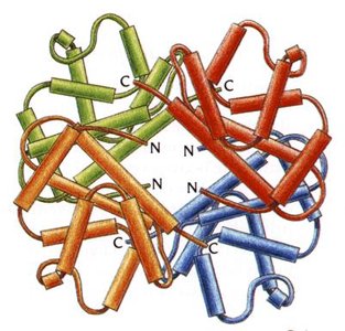
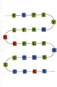
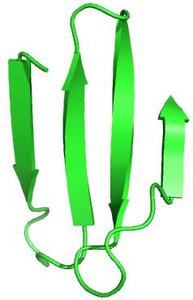
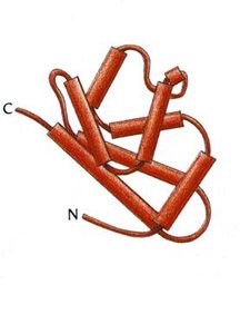
Proteins are complex biomolecules whose function is determined by their structure. The structure of proteins is organized into four hierarchical levels: primary, secondary, tertiary, and quaternary.
Primary Structure: The sequence of amino acids in a polypeptide chain, held together by peptide bonds.
Secondary Structure: Local folding patterns such as alpha helices and beta sheets, stabilized by hydrogen bonds.
Tertiary Structure: The overall three-dimensional shape of a single polypeptide chain, stabilized by interactions among side chains.
Quaternary Structure: The arrangement of multiple polypeptide chains (subunits) into a functional protein complex.
Shape Determining Interactions
The folding and stability of proteins are governed by several types of interactions:
Covalent Bonds: Intramolecular bonds such as peptide bonds and disulfide bridges.
Electrostatic Interactions (Salt Bridges): Ionic bonds between acidic and basic side chains.
Hydrogen Bonds: Occur between hydrogen bond donors (O-H, N-H) and acceptors (O, N).
Van der Waals Forces: Weak interactions between all atoms, important for close packing.
Classification of Protein Structures
Proteins are classified based on their shape and solubility:
Globular Proteins: Globe-like, typically water soluble due to hydrophilic residues on the surface. Examples include enzymes and transport proteins.
Fibrous Proteins: Form fibers or sheets, typically insoluble, with tightly packed hydrophobic residues. Examples include hair, teeth, and fingernails.
Conjugated Proteins
Types of Conjugated Proteins
Conjugated proteins require non-protein components for their function. The main types include:
Glycoproteins: Covalently bound carbohydrates (e.g., cell surface receptors).
Lipoproteins: Covalently bound lipids (e.g., cholesterol transporters).
Metalloproteins: Contain metal ions for structural or catalytic functions (e.g., zinc-stabilized proteins).
Phosphoproteins: Covalently bound phosphate groups for activation/deactivation or binding sites.
Hemoproteins: Contain heme groups/porphyrin rings (e.g., oxygen transporters).
Nucleoproteins: Contain nucleic acids (e.g., ribosomes).
Myoglobin and Hemoglobin: Structure and Function
Myoglobin
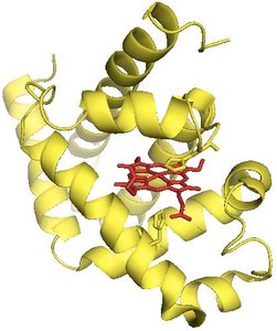
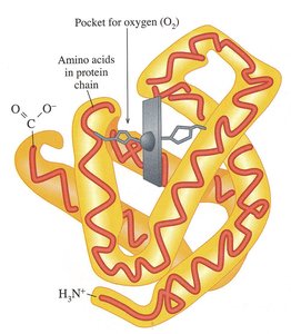
Myoglobin is a globular protein found in muscle tissue. It consists of a single polypeptide chain (tertiary structure) and functions as an oxygen storage molecule.
Structure: Tertiary, single polypeptide chain.
Function: Stores oxygen in muscle cells.
Location: Muscles.
Hemoglobin
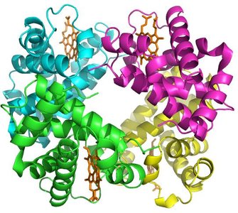
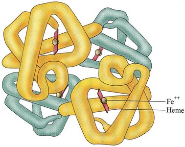
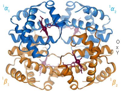
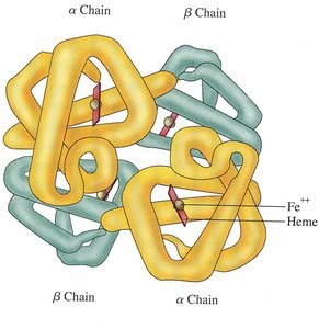
Hemoglobin is a globular protein found in red blood cells. It has a quaternary structure, consisting of four polypeptide chains (two alpha and two beta), and functions as an oxygen transporter.
Structure: Quaternary, four polypeptide chains (2 alpha, 2 beta).
Function: Transports oxygen from lungs to tissues.
Location: Red blood cells.
Oxygen Transport and Binding
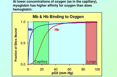
Hemoglobin and myoglobin differ in their oxygen binding properties. Hemoglobin exhibits cooperative binding, while myoglobin binds oxygen with higher affinity at low concentrations.
Hemoglobin: Cooperative binding, releases oxygen in tissues.
Myoglobin: High affinity for oxygen, stores oxygen in muscle.
Sickle Cell Anemia: Structural and Functional Changes
Physical Changes in Hemoglobin
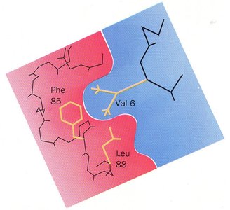
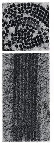
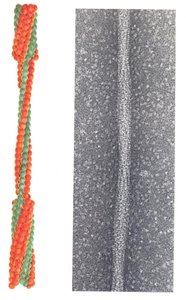
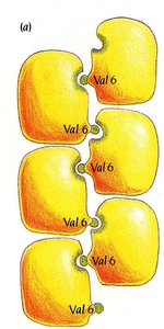
Sickle cell anemia is caused by a mutation in the hemoglobin gene, resulting in the substitution of glutamic acid (hydrophilic) with valine (hydrophobic) at position 6 of the beta chain. This change promotes aggregation of hemoglobin molecules, distorting red blood cells into a sickle shape.
Mutation: Glu → Val at position 6 in beta chain.
Effect: Hydrophobic valine interacts with hydrophobic pockets, causing aggregation.
Symptoms: Anemia, pain, fatigue, stroke.
Electrophoresis: Analytical and Diagnostic Tool
Principles of Electrophoresis
Electrophoresis is a technique used to separate proteins based on their charge and molecular weight. It is commonly used to diagnose sickle cell anemia by distinguishing normal hemoglobin (HbA) from sickle cell hemoglobin (HbS).
Separation: Proteins migrate in an electric field according to their charge and size.
Diagnostic Use: HbA migrates closer to the anode than HbS due to charge differences.
Collagen: Structure, Function, and Related Diseases
Collagen Structure and Function
Collagen is a fibrous protein with a quaternary structure, found in connective tissues such as bones, teeth, skin, and tendons. Its structure is stabilized by hydrogen bonds and covalent cross-links.
Subunit: Tropocollagen, composed of three polypeptide chains.
Stabilization: Hydrogen bonds and covalent (disulfide) bonds.
Function: Provides structural support and strength to tissues.
Collagen Related Diseases
Defects in collagen synthesis or structure can lead to diseases such as osteogenesis imperfecta (brittle bone disease) and scurvy (vitamin C deficiency).
Brittle Bone Disease: Insufficient or poor quality collagen, leading to fractures and weak tissues.
Scurvy: Lack of vitamin C impairs hydroxylation of proline, resulting in defective collagen.
Keratin: Structure and Stabilization
Alpha and Beta Keratin
Keratin is a fibrous protein found in hair, nails, and skin. Alpha-keratin is stabilized by disulfide bonds, while beta-keratin is stabilized by van der Waals interactions.
Alpha-Keratin: Disulfide bonds between peptide chains provide strength and stability.
Beta-Keratin: Van der Waals interactions stabilize the packing of strands.
Protein Folding and Chaperones
Chaperones
Chaperones are proteins and protein complexes that assist in the folding of other proteins, preventing aggregation and ensuring proper function.
Function: Assist in protein folding, prevent aggregation.
Importance: Essential for cellular health and prevention of misfolded protein diseases.
Prions: Misfolded Infectious Proteins
Prion Diseases
Prions are misfolded proteins that can induce other proteins to misfold, leading to transmissible spongiform encephalopathies (TSEs), which are fatal degenerative brain disorders.
Mechanism: Conversion of alpha-helices to beta-sheets, inducing misfolding in other proteins.
Transmission: Through infected tissue, body fluids, or contaminated instruments.
Diseases: TSEs, including Creutzfeldt-Jakob disease and mad cow disease.
Protein Degradation and Denaturation
Protein Degradation
Protein degradation is the breakdown of proteins into their constituent amino acids. It occurs via hydrolysis of peptide bonds and is facilitated by lysosomes, proteasomes, and digestive enzymes.
Hydrolysis: Peptide bonds are broken, releasing amino acids.
Methods: Autophagy, lysosomal degradation, proteasomal degradation.
Protein Denaturation
Protein denaturation is the unfolding of proteins, resulting in loss of function. It is caused by physical or chemical agents such as heat, mechanical agitation, detergents, organic solvents, pH changes, and inorganic salts.
Physical Process: Disrupts non-covalent interactions, leading to loss of structure and function.
Agents: Heat, agitation, detergents, solvents, pH, salts.
Summary Table: Protein Structure Levels
Level | Description | Bond/Interaction |
|---|---|---|
Primary | Amino acid sequence | Peptide bonds |
Secondary | Alpha helices, beta sheets | Hydrogen bonds |
Tertiary | 3D folding of single chain | Hydrogen bonds, ionic bonds, van der Waals, disulfide bonds |
Quaternary | Assembly of multiple chains | Same as tertiary, between chains |
Additional info: These notes expand on the lecture outline, providing definitions, examples, and structural context for protein function and disease. Images included are directly relevant to the structural and functional explanations of proteins.
