 Back
Protein Structure and Function: Tertiary, Quaternary Structure, and Disease
Study Guide - Smart Notes
 Tailored notes based on your materials, expanded with key definitions, examples, and context.
Protein Structure and Function
Levels of Protein Structure
Proteins are complex biomolecules whose function is determined by their structure. The structure of proteins is organized into four hierarchical levels: primary, secondary, tertiary, and quaternary.
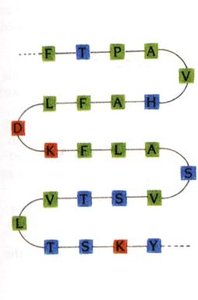
Primary Structure: The linear sequence of amino acids in a polypeptide chain, held together by peptide bonds.
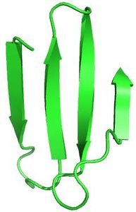
Secondary Structure: Local folding patterns such as alpha-helices and beta-sheets, stabilized by hydrogen bonds.
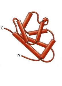
Tertiary Structure: The overall three-dimensional shape of a single polypeptide chain, stabilized by interactions among side chains (R groups).
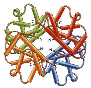
Quaternary Structure: The arrangement of multiple polypeptide chains (subunits) in a functional protein complex.
Shape-Determining Interactions
The final shape of a protein is determined by several types of interactions:
Covalent Bonds: Such as disulfide bonds between cysteine residues, which stabilize the structure.
Electrostatic Interactions (Salt Bridges): Ionic bonds between acidic and basic side chains.
Hydrogen Bonds: Occur between hydrogen donors (O-H, N-H) and acceptors (O, N).
Van der Waals Forces: Weak attractions between all atoms in close proximity.
Classification of Protein Structures
Globular Proteins: Compact, globe-like, typically water-soluble due to hydrophilic residues on the surface. Examples: enzymes, myoglobin, hemoglobin.
Fibrous Proteins: Extended, fiber-like, typically insoluble, with hydrophobic residues packed together. Examples: collagen, keratin.
Conjugated Proteins
Definition and Types
Conjugated proteins require non-protein components (prosthetic groups) for their function. The main types include:
Glycoproteins: Contain covalently bound carbohydrates (e.g., cell surface receptors).
Lipoproteins: Bound to lipids (e.g., cholesterol transporters).
Metalloproteins: Require metal ions for structure or catalysis (e.g., zinc-containing enzymes).
Phosphoproteins: Contain covalently bound phosphate groups (e.g., regulatory proteins).
Hemoproteins: Contain heme groups (e.g., hemoglobin, myoglobin).
Nucleoproteins: Contain nucleic acids (e.g., ribosomes).
Oxygen Transport Proteins: Myoglobin and Hemoglobin
Myoglobin
Myoglobin is a globular, conjugated protein found in muscle tissue. It consists of a single polypeptide chain (tertiary structure) and binds oxygen for storage and release during muscle activity.
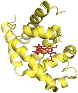
Location: Muscle cells
Function: Oxygen storage and release
Structure: Single polypeptide chain with a heme prosthetic group
Hemoglobin
Hemoglobin is a globular, conjugated protein found in red blood cells. It has a quaternary structure, consisting of four polypeptide chains (two alpha and two beta subunits), each with a heme group for oxygen binding and transport.
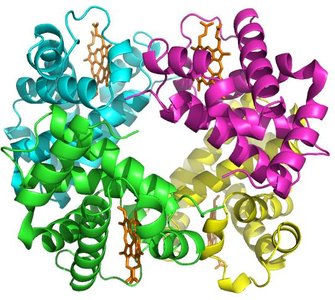
Location: Red blood cells
Function: Oxygen transport from lungs to tissues
Structure: Four polypeptide chains (2α, 2β) with heme groups
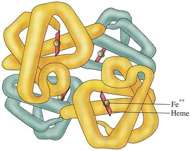
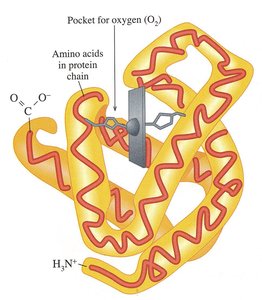
Oxygen Binding and Transfer
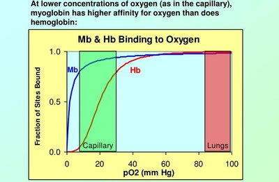
Hemoglobin and myoglobin differ in their oxygen-binding properties. Myoglobin has a higher affinity for oxygen at low concentrations, facilitating oxygen transfer from hemoglobin to myoglobin in muscle tissue.
Sickle Cell Anemia: Structure and Disease
Molecular Basis of Sickle Cell Anemia
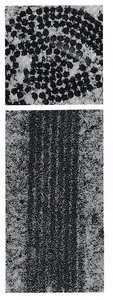
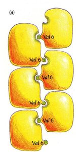
Sickle cell anemia is caused by a single amino acid substitution (Glu → Val at position 6) in the beta chain of hemoglobin. This change introduces a hydrophobic residue, leading to abnormal aggregation of hemoglobin molecules under low oxygen conditions.
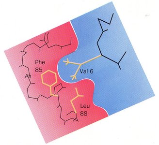
Normal Hemoglobin (HbA): Glutamic acid at position 6 (hydrophilic)
Sickle Cell Hemoglobin (HbS): Valine at position 6 (hydrophobic)
Effect: Aggregation of hemoglobin, deformation of red blood cells, reduced oxygen transport
Electrophoresis in Protein Analysis
Electrophoresis is a laboratory technique used to separate proteins based on their charge and size. It is commonly used to distinguish between normal and sickle cell hemoglobin due to their different charges resulting from the amino acid substitution.
Fibrous Proteins: Collagen and Keratin
Collagen
Collagen is a fibrous protein with a quaternary structure, found in connective tissues such as bone, skin, and tendons. It consists of three polypeptide chains wound into a triple helix, stabilized by hydrogen bonds and covalent cross-links.
Subunit: Tropocollagen
Key Residues: Glycine (every third residue), proline, and hydroxyproline
Function: Provides structural strength and flexibility
Collagen-Related Diseases
Brittle Bone Disease (Osteogenesis Imperfecta): Caused by insufficient or defective collagen.
Scurvy: Caused by vitamin C deficiency, leading to defective collagen synthesis.
Keratin
Keratin is a fibrous protein found in hair, nails, and skin. Its structure is stabilized by a high number of disulfide bonds, which provide strength and resilience.
Alpha-Keratin: Forms coiled coils stabilized by disulfide bonds.
Beta-Keratin: Stabilized by van der Waals interactions.
Protein Folding, Chaperones, and Misfolding
Chaperones
Chaperones are proteins that assist in the proper folding of other proteins, preventing aggregation and misfolding.
Prions and Infectious Protein Diseases
Prions are misfolded proteins that can induce normal proteins to adopt the abnormal conformation, leading to transmissible spongiform encephalopathies (TSEs), a group of fatal neurodegenerative diseases.
Protein Degradation and Denaturation
Protein Degradation
Protein degradation is the process by which proteins are broken down into their constituent amino acids. This occurs via hydrolysis of peptide bonds, primarily in lysosomes, proteasomes, the stomach, and the small intestine.
Autophagy: Cellular process for degrading and recycling cellular components.
Lysosomes: Organelles that digest unwanted proteins and organelles.
Proteasomes: Degrade unfolded or unneeded proteins by proteolysis.
Protein Denaturation
Denaturation is the loss of a protein's native structure due to physical or chemical agents, resulting in loss of function. It is primarily a physical process and can be caused by heat, pH changes, detergents, organic solvents, or mechanical agitation.
Heat: Disrupts non-covalent interactions.
pH Change: Alters charge on side chains, disrupting salt bridges.
Detergents: Interact with hydrophobic regions, unfolding the protein.
Organic Compounds: Disrupt hydrogen bonds.
Summary Table: Key Protein Types and Features
Protein | Structure | Location | Function | Key Feature |
|---|---|---|---|---|
Myoglobin | Tertiary (single chain) | Muscle | Oxygen storage | Heme group |
Hemoglobin | Quaternary (4 chains) | Red blood cells | Oxygen transport | Heme groups, cooperative binding |
Collagen | Quaternary (triple helix) | Connective tissue | Structural support | Glycine, proline, hydroxyproline |
Keratin | Fibrous (coiled coil) | Hair, nails, skin | Protection, structure | Disulfide bonds |
Key Equations
Peptide Bond Hydrolysis:
Oxygen Binding (Hemoglobin):
Additional info: This summary integrates foundational concepts from GOB Chemistry Chapter 18 (Amino Acids and Proteins), focusing on protein structure, function, and disease relevance.
