 Back
Radiation Biology: Mechanisms, Effects, and Measurement in GOB Chemistry
Study Guide - Smart Notes
 Tailored notes based on your materials, expanded with key definitions, examples, and context.
Radiation Biology
Introduction to Radiation Injury
Radiation biology examines the effects of ionizing radiation on living tissues, focusing on the mechanisms of injury, dose-response relationships, and the risks associated with exposure. Understanding these principles is essential for evaluating the safety and risks of medical and dental radiographic procedures.
Key Terms and Mechanisms of Radiation Injury
Ionizing Radiation: Radiation with enough energy to remove tightly bound electrons from atoms, thus creating ions.
Ionization: The process by which an atom or molecule acquires a positive or negative charge by gaining or losing electrons.
Free Radicals: Highly reactive and unstable atoms or molecules with unpaired electrons, often formed when radiation ionizes water in cells.
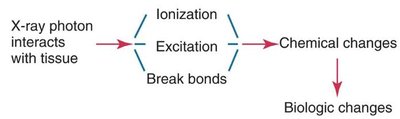
When x-ray photons interact with tissue, they may cause ionization, excitation, or break chemical bonds, leading to chemical and ultimately biologic changes.
Mechanisms of Injury: Ionization and Free Radical Formation
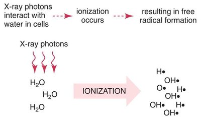
Ionization can occur via the photoelectric effect or Compton scatter, resulting in a positive atom and a dislodged electron. This electron can further react with other atoms, causing biological damage. Most cell damage from radiation occurs through the formation of free radicals when x-ray photons ionize water molecules in cells.
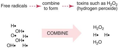
Free radicals can combine to form toxins such as hydrogen peroxide (H2O2), which are harmful to cells.
Theories of Radiation Injury: Direct and Indirect Effects
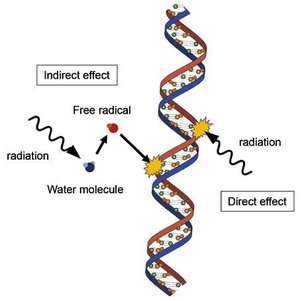
There are two main theories describing how radiation damages biological tissues:
Direct Theory: Radiation directly hits a critical part of the cell, such as DNA, causing damage. This occurs infrequently.
Indirect Theory: Radiation is absorbed by water in the cell, forming free radicals that combine into toxins, which then damage the cell. This is the more common mechanism.
Dose-Response Curve
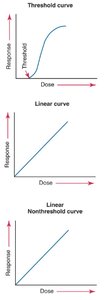
The dose-response curve correlates the amount of radiation received with the extent of tissue damage. The relationship can be:
Threshold Curve: No response is seen below a certain dose; above this threshold, response increases with dose.
Linear Curve: Response is directly proportional to dose.
Linear Nonthreshold Curve: Any dose, no matter how small, can cause a response; risk increases linearly with dose.
Stochastic and Nonstochastic (Deterministic) Effects
Stochastic Effects: Probability of occurrence increases with dose, but severity does not. No threshold; examples include cancer and genetic mutations.
Nonstochastic (Deterministic) Effects: Severity increases with dose and a threshold exists. Examples include erythema, hair loss, cataracts, and decreased fertility.
Sequence of Radiation Injury
Latent Period: Time between exposure and appearance of observable clinical signs; depends on total dose and rate.
Period of Injury: Cellular injuries manifest.
Recovery Period: Cells may repair damage, depending on various factors.
Cumulative Effects: Unrepaired damage accumulates, increasing risk over time.
Short-Term and Long-Term Effects
Short-Term Effects: Result from large doses in a short time (e.g., acute radiation syndrome: nausea, vomiting, diarrhea, hair loss, hemorrhage).
Long-Term Effects: Result from small doses absorbed repeatedly over a long period (e.g., cancer, birth abnormalities, genetic defects).
Somatic and Genetic Effects
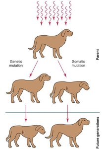
Somatic Effects: Affect all cells except reproductive cells; seen in the irradiated individual, not passed to offspring.
Genetic Effects: Affect reproductive cells; not seen in the irradiated individual but passed to future generations.
Radiation Effects on Cells, Tissues, and Organs
Mitotic Activity: Cells that divide frequently are more sensitive to radiation.
Cell Differentiation: Immature or unspecialized cells are more sensitive.
Cell Metabolism: Cells with higher metabolism are more sensitive.
Radiosensitive tissues include bone marrow, reproductive cells, and intestines. Radioresistant tissues include salivary glands, kidney, and liver. Critical organs exposed during dental radiography include the skin, thyroid gland, lens of the eye, and bone marrow.
Units of Measurement in Radiation Exposure
Traditional Units: Roentgen (R), rad (radiation absorbed dose), rem (roentgen equivalent man).
SI Units: Coulombs/kilogram (C/kg), gray (Gy), sievert (Sv).
Key conversions:
1 R = 1 C/kg
100 rad = 1 Gy
100 rem = 1 Sv
For dental imaging, doses are often expressed in millisieverts (mSv, 1/1000 Sv) or microsieverts (µSv, 1/1,000,000 Sv).
Sources of Radiation Exposure
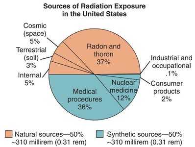
Radiation exposure comes from both natural and artificial sources:
Natural Background Radiation: Cosmic (space), terrestrial (soil), internal (within the body), and radon/thoron gases.
Artificial (Humanmade) Radiation: Medical procedures, consumer products, fallout from atomic weapons, and the nuclear fuel cycle.
Risk and Risk Estimates for Radiation Exposure
The risk of dental imaging inducing a fatal cancer is extremely low (about 3 in 1,000,000), especially compared to other everyday risks. For example, the risk of developing cancer spontaneously is much higher (3300 in 1,000,000).
Dental Radiation and Exposure Risks
Thyroid Gland: Not directly irradiated in dental imaging; cancer risk requires much higher doses than those used in dentistry.
Bone Marrow: Only a small percentage is exposed during dental imaging; leukemia risk is negligible at dental doses.
Skin: Erythema requires much higher doses than those encountered in dental radiography.
Eyes: Cataract formation requires doses far exceeding those used in dental imaging.
Patient Exposure and Dose Reduction
Receptor Choice: Digital sensors reduce radiation exposure by up to 50% compared to conventional film.
Collimation: Rectangular collimation reduces absorbed dose by up to 60% compared to round collimation.
Technique: Increasing the target-receptor distance and using the paralleling technique reduces skin dose.
Risk Versus Benefit of Dental Images
Dental images should only be prescribed when the benefit of disease detection outweighs the risk of biologic damage. When properly prescribed and exposed, the benefits far outweigh the risks.
Natural Radioactivity in Food
Trace radionuclides such as potassium, carbon, and radium occur naturally in food. For example, bananas are high in potassium and contribute about 0.1 µSv per banana, which is negligible compared to daily background exposure.
Comparisons of Radiation Doses
The following table compares the dose equivalents from various sources, including dental x-rays, medical procedures, and natural background radiation.
Radiation Source | Dose Equivalent (mSv) |
|---|---|
Max annual dose permitted for U.S. radiation workers | 50 |
Whole body CT scan | 10 |
Abdominal CT scan | 6-10 |
Chest CT scan | 6 |
Average annual dose from natural sources (U.S.) | 2.8 |
Head CT scan | 2 |
Annual dose from food and water (potassium) | 0.4 |
Smoking one pack of cigarettes per day (annual) | 0.36 |
Dental panoramic digital | 0.014-0.024 |
Dental bitewing digital | 0.005 |
Airport security scan | 0.0001 |
Living in a brick/concrete house (annual) | 0.007 |
Additional info: Table entries inferred from the referenced chart and typical values for dental and medical exposures. |
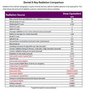
Example: A dental bitewing x-ray delivers about 0.005 mSv, which is a tiny fraction of the annual background radiation dose.
