 Back
Blood: Structure, Function, and Homeostasis in Human Biology
Study Guide - Smart Notes
 Tailored notes based on your materials, expanded with key definitions, examples, and context.
The Circulatory System
Overview and Functions
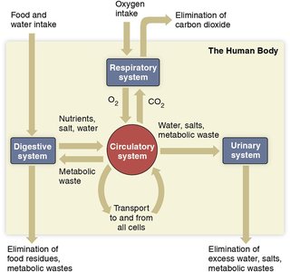
The circulatory system is a vital organ system responsible for transporting materials throughout the body, maintaining homeostasis, and defending against disease and injury. Blood is the fluid medium within this system, interacting with other organ systems to ensure proper physiological function.
Transport: Delivers nutrients, oxygen, and hormones to cells; removes waste products.
Regulation: Maintains body temperature, water balance, and pH.
Defense: Protects against pathogens and minimizes blood loss after injury.
Blood: A Specialized Connective Tissue
Composition and Properties
Blood is a specialized connective tissue composed of cells and cell fragments suspended in plasma. It originates from bone marrow and constitutes about 8% of body weight.
Volume: Adult men average 5-6L; women 4-5L.
Uniformity: Blood is uniform in color but contains components of varying densities.
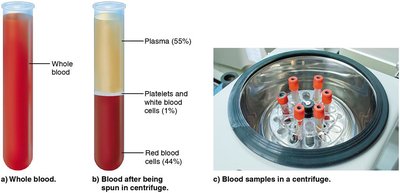
Components: Blood consists of formed elements (cells and cell fragments) and plasma (liquid portion).
Blood Composition
Formed Elements (45%)
Formed elements include red blood cells (RBCs), white blood cells (WBCs), and platelets.
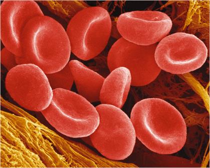
Red Blood Cells (Erythrocytes): Transport oxygen (O2) and carbon dioxide (CO2).
White Blood Cells (Leukocytes): Defend against pathogens and abnormal cells.
Platelets: Essential for blood clotting.
Plasma (55%)
Plasma is the pale yellow, liquid portion of blood, carrying cells and platelets.
Water: Main component.
Electrolytes: Na, K, Cl, HCO3, Ca, H, Mg regulate cell function and excitability.
Proteins: Albumins, globulins, clotting proteins.
Hormones: Chemical messengers for regulation.
Gases: O2 for metabolism, CO2 as waste.
Nutrients & Wastes: Glucose, urea, and other materials transported throughout the body.
Plasma Proteins
Types and Functions
Plasma proteins play critical roles in maintaining homeostasis and supporting physiological functions.
Albumins: Maintain water balance and transport drugs/molecules.
Globulins: Transport substances; beta globulins form lipoproteins (LDL, HDL); gamma globulins fight infection.
Clotting Proteins: Essential for blood clotting and minimizing blood loss.
Red Blood Cells (RBCs)
Structure and Function
RBCs, or erythrocytes, are highly specialized for gas transport.
Shape: Flexible, biconcave discs allow passage through tiny vessels.
Design: Mature RBCs lack a nucleus and organelles; contain ~300 million hemoglobin molecules.
Function: Carry O2 and CO2; give blood its red color and viscosity.
Hemoglobin
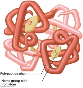
Hemoglobin is the O2-binding protein in RBCs, consisting of four polypeptide chains, each with a heme group containing an iron atom.
Oxygen Binding: O2 binds to iron in heme groups; one RBC can carry up to 1.2 billion O2 molecules.
ATP Production: RBCs produce ATP anaerobically (no mitochondria).
Hemoglobin & Oxygen Binding
Binding: Occurs in lungs where O2 concentration and pH are high.
Oxyhemoglobin: Temporary bond between hemoglobin and O2.
Release: Occurs in tissues with lower O2 concentration, lower pH, or increased heat.
Deoxyhemoglobin: Hemoglobin without O2.
CO2 Transport: Hemoglobin also carries CO2 at a different site.
Hematocrit
Definition and Clinical Significance
Hematocrit is the percentage of blood volume occupied by RBCs, reflecting oxygen-carrying capacity.
Normal Ranges: Men: 43-49%; Women: 37-43%.
Low Hematocrit: Indicates anemia or low RBC production.
High Hematocrit: Indicates excessive RBC production, risk of clots.
Altitude: Hematocrit rises at higher altitudes as a homeostatic adjustment.
Blood Cell Production
Stem Cells and Differentiation
All blood cells and platelets originate from stem cells in red bone marrow.
Stem Cells: Unspecialized cells producing immature "blasts" that mature into blood cells and platelets.
Erythroblasts: Immature RBCs that fill with hemoglobin and lose their nucleus.
Megakaryoblasts: Immature cells that mature into megakaryocytes, which fragment to form platelets.
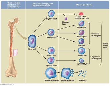
Red Blood Cell & Platelet Formation
Process and Lifespan
Erythrocytes: Mature RBCs live ~120 days, produced at a rate of 2 million per second.
Megakaryocytes: Large cells with multi-lobular nuclei; fragmentation produces platelets (lifespan 5-9 days).
RBC Lifespan and Recycling
Removal and Breakdown
Old and damaged RBCs are removed by macrophages in the liver and spleen, which engulf and digest them (phagocytosis).
Peptide Chains: Broken down into amino acids for new protein synthesis.
Iron: Returned to bone marrow for reuse in hemoglobin.
Heme Group: Converted to bilirubin, a yellow pigment excreted in bile.
Bilirubin and Jaundice
Bilirubin: Contributes to yellow color of urine and feces; color change in bruising.
Jaundice: Yellowing of skin and eyes due to improper bilirubin secretion by the liver.
Regulation of RBC Production
Erythropoietin and Negative Feedback
RBC production is regulated by the hormone erythropoietin, released by the kidneys in response to low blood oxygen levels.
Erythropoietin: Stimulates stem cells in bone marrow to produce more RBCs.
Negative Feedback: When O2 levels normalize, erythropoietin release decreases.
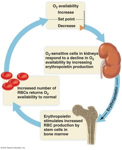
Blood Doping
Artificial Erythropoietin: Used by athletes to increase oxygen-carrying capacity.
Risks: Increased blood viscosity, heart strain, risk of clots, high blood pressure, heart attack, and stroke.
Platelet Production and Regulation
Thrombopoietin
Platelet production is regulated by thrombopoietin, a hormone produced in the liver and kidneys.
Platelets: Small cell fragments essential for blood clotting.
Thrombopoietin: Stimulates platelet formation in bone marrow.
White Blood Cells (WBCs)
Types and Functions
WBCs, or leukocytes, are larger and more diverse than RBCs, working with the lymphatic system to defend against pathogens.
Granular Leukocytes (Granulocytes): Neutrophils, eosinophils, basophils (lifespan: hours to 9 days).
Agranular Leukocytes (Agranulocytes): Monocytes (months), lymphocytes (days to years; B- and T-lymphocytes).
Structure: Have a nucleus, lack hemoglobin; only 1 WBC per 700 RBCs.
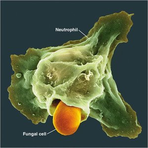
Hemostasis: Stopping Blood Loss
Stages of Hemostasis
Hemostasis is the natural process of stopping blood loss, involving three stages:
Vascular Spasms: Smooth muscle contraction constricts blood vessels for ~30 minutes.
Platelet Plug: Platelets become sticky and clump together, sealing the injury.
Blood Clotting: 12 clotting factors, including prothrombin activator, thrombin, and fibrinogen, work together to reduce blood flow.
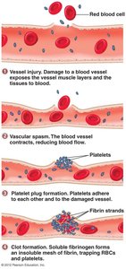
Blood Clotting Mechanism
Prothrombin Activator: Converts prothrombin to thrombin.
Thrombin: Converts fibrinogen to fibrin.
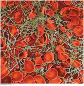
Fibrin: Forms threads that create a mesh, trapping platelets and blood cells to form a clot.
Clot: Reduces blood flow and tightens vessel walls.
Summary Table: Blood Components
Component | Function | Percentage |
|---|---|---|
Red Blood Cells (RBCs) | Transport O2 and CO2 | ~44% |
White Blood Cells (WBCs) | Defense against pathogens | ~1% |
Platelets | Blood clotting | ~1% |
Plasma | Transport nutrients, wastes, hormones | ~55% |
Key Terms and Definitions
Hemoglobin: O2-binding protein in RBCs.
Hematocrit: Percentage of blood volume occupied by RBCs.
Erythropoietin: Hormone regulating RBC production.
Thrombopoietin: Hormone regulating platelet production.
Hemostasis: Process of stopping blood loss.
Bilirubin: Yellow pigment from heme breakdown.
Relevant Equations
Hematocrit Calculation
Hematocrit (%) =
Oxygen Transport
O2 + Hemoglobin Oxyhemoglobin
Blood Clotting Cascade
Prothrombin Thrombin
Fibrinogen Fibrin
Example: Blood Doping in Athletics
Athletes may use synthetic erythropoietin to increase RBC count and oxygen-carrying capacity, but this poses significant health risks.
Additional info:
Blood cell production and regulation are tightly controlled by feedback mechanisms to maintain homeostasis.
Blood composition and function are central to understanding human physiology and disease.
