 Back
Bone Types and Structures: Anatomy and Cellular Organization of Long Bones
Study Guide - Smart Notes
 Tailored notes based on your materials, expanded with key definitions, examples, and context.
The Skeletal System
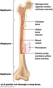
Long Bone Structure
Long bones are a fundamental component of the human skeletal system, characterized by their elongated shape and specialized internal architecture. Understanding the anatomy of a long bone is essential for comprehending its function in support, movement, and hematopoiesis.
Diaphysis: The cylindrical shaft of the bone, composed primarily of compact bone, providing strength and support.
Epiphysis: The rounded ends of the bone, containing spongy bone filled with red bone marrow and stem cells, crucial for blood cell production.
Compact Bone: Dense, solid bone tissue forming the outer layer, offering protection and structural integrity.
Spongy Bone: Porous bone tissue within the epiphysis, housing red bone marrow.
Periosteum: Tough connective tissue covering the bone's surface, containing bone-forming cells (osteocytes).
Central Cavity: Located within the diaphysis, filled with yellow bone marrow, which stores fat.
Blood Vessels: Supply nutrients and oxygen to bone cells via the central canal.
Microscopic Structure of Bone: Osteons and Osteocytes
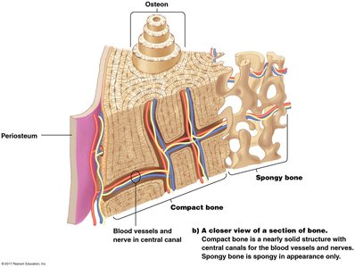
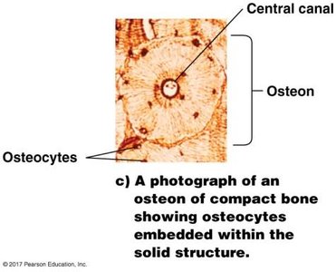
Bone tissue is organized into cylindrical structures called osteons, which are essential for nutrient delivery and waste removal. Osteocytes, the mature bone cells, are embedded within the solid matrix and maintain bone health.
Osteon: The fundamental unit of compact bone, consisting of concentric rings (lamellae) surrounding a central canal (Haversian canal).
Haversian Canal: Central channel within each osteon, containing blood vessels and nerves.
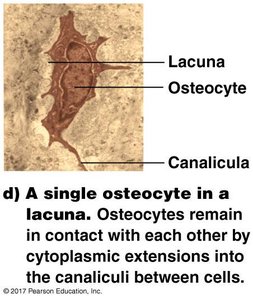
Osteocytes: Bone cells trapped within small chambers (lacunae) in the matrix; they maintain bone tissue.
Lacunae: Small cavities housing osteocytes.
Canaliculi: Tiny canals connecting lacunae, allowing communication and nutrient exchange between osteocytes.
Gap Junctions: Specialized connections facilitating direct transfer of nutrients and signals between osteocytes.
Cellular Communication and Bone Hardening
As bone develops, osteocytes become enclosed by calcium phosphate, which hardens the matrix. The canaliculi and gap junctions ensure that even isolated cells can exchange nutrients and maintain bone health.
Bone Hardening: Calcium phosphate deposition leads to a rigid structure.
Cellular Contact: Osteocytes remain interconnected via canaliculi, supporting tissue maintenance.
Summary Table: Key Structures of a Long Bone
Structure | Location | Function |
|---|---|---|
Diaphysis | Shaft | Support, strength |
Epiphysis | Ends | Blood cell production, joint articulation |
Compact Bone | Outer layer | Protection, structure |
Spongy Bone | Epiphysis interior | Red marrow storage |
Periosteum | Surface | Bone growth, repair |
Osteon | Compact bone | Nutrient delivery |
Osteocyte | Lacunae | Bone maintenance |
Canaliculi | Between lacunae | Cell communication |
Additional info:
Long bones are essential for movement and leverage in the body, and their internal structure is optimized for both strength and metabolic activity. The arrangement of osteocytes and osteons allows for efficient nutrient exchange and adaptation to mechanical stress.
