 Back
Chapter 9: The Immune System and Mechanisms of Defense – Study Notes
Study Guide - Smart Notes
 Tailored notes based on your materials, expanded with key definitions, examples, and context.
The Immune System and Mechanisms of Defense
Overview of the Body’s Defense Mechanisms
The human body employs multiple defense mechanisms to protect against pathogens. These mechanisms are categorized as barriers, nonspecific defenses, and specific immune responses.
Barriers to Entry: Physical and chemical barriers such as skin, stomach acid, tears, and vomiting prevent pathogens from entering or neutralize them.
Nonspecific Defense Mechanisms: Includes phagocytosis and inflammation, which act against a wide range of pathogens.
Specific Defense Mechanisms: The immune response targets specific pathogens using antibodies and T cells.
Pathogens Cause Disease
Pathogens are disease-causing agents that can be living organisms or nonliving infectious particles.
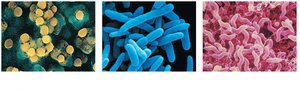
Bacteria: Unicellular prokaryotes causing diseases like pneumonia and tuberculosis.
Fungi: Unicellular or multicellular eukaryotes.
Parasites: Unicellular or multicellular eukaryotes.
Viruses: Nonliving infectious particles.
Prions: Infectious proteins causing neurodegenerative diseases.
Cellular Structure of Pathogens
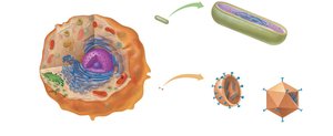
Bacteria, viruses, and eukaryotic cells differ in structure and complexity.
Bacterial Cells: Prokaryotic, with a single strand of DNA and free-floating ribosomes. Surrounded by a rigid cell wall.
Eukaryotic Cells: Have a membrane-bound nucleus and organelles.
Viruses: Consist of a protein coat surrounding either RNA or DNA.
Bacteria: Single-Celled Living Organisms
Bacteria are prokaryotic, single-celled organisms that utilize various resources for growth and reproduction. They cause a range of infections and are generally treated with antibiotics.
Examples of Bacterial Infections: Pneumonia, tonsillitis, tuberculosis, botulism, syphilis, Lyme disease.
Treatment: Antibiotics target bacterial cells.
Viruses: Tiny Infectious Agents
Viruses are much smaller than bacteria and are not considered living. They require a host cell to reproduce and lack metabolic activity.
Structure: DNA or RNA surrounded by a protein coat (capsid).
Diseases: AIDS, hepatitis, influenza, colds, chicken pox, Ebola.
Prions: Infectious Proteins
Prions are misfolded proteins that propagate by inducing misfolding in normal proteins, leading to cell dysfunction.
Resistant to: Cooking, freezing, drying.
Diseases: Bovine spongiform encephalitis (mad cow disease), variant Creutzfeldt-Jakob disease.
Transmissibility, Mode of Transmission, and Virulence
The health risk posed by a pathogen depends on how easily it spreads (transmissibility), its mode of transmission, and its virulence (damage caused).
Modes of Transmission: Respiratory, fecal–oral, body fluids, direct contact.
Virulence: Severity of infection.
The Lymphatic System: Defense and Support
Functions of the Lymphatic System
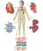
The lymphatic system plays a crucial role in defending the body against infection, maintaining blood volume, and transporting fats.
Maintenance of Blood Volume: Supports cardiovascular function.
Transport of Fats: Moves fats from the digestive system to the cardiovascular system.
Filtration: Removes foreign material to defend against infection.
Lymphatic Vessels and Lymph
Lymphatic vessels form a network similar to veins, beginning as blind-ended capillaries and draining into the cardiovascular system. Lymph is a milky fluid containing white blood cells, proteins, fats, and sometimes pathogens.
Lymph Nodes
Lymph nodes are small structures that filter lymph, removing microorganisms, debris, and abnormal cells. They are composed of connective tissue, macrophages, and lymphocytes.
The Spleen
The spleen is the largest lymphatic organ, located in the upper left abdominal cavity. It removes old and damaged red blood cells and microorganisms from blood and contains lymphocytes searching for pathogens.
Thymus Gland, Tonsils, and Adenoids
The thymus gland is the site of T cell maturation and secretes hormones for T cell development. Tonsils and adenoids filter food and air entering the throat and nasal passages, respectively.
Body’s First Line of Defense: Physical and Chemical Barriers
Physical and Chemical Barriers
The first line of defense includes skin, tears, saliva, ear wax, mucus, stomach acid, vaginal acidity, vomiting, urination, defecation, and resident bacteria.
Lysozyme: An antibacterial enzyme found in tears and saliva.
Resident Bacteria: Outcompete pathogens for resources.
Nonspecific Defenses: Second Line of Defense
Components of Nonspecific Defense
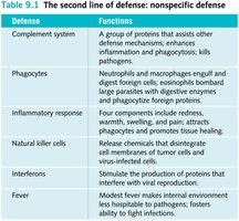
Nonspecific defenses act against a broad range of pathogens and include phagocytic cells, inflammation, natural killer cells, complement proteins, interferons, and fever.
Defense | Functions |
|---|---|
Complement system | Assists other defense mechanisms; enhances inflammation and phagocytosis; kills pathogens. |
Phagocytes | Engulf and digest foreign cells; neutrophils and macrophages are key types. |
Inflammatory response | Redness, warmth, swelling, pain; attracts phagocytes and promotes healing. |
Natural killer cells | Disintegrate cell membranes of tumor and virus-infected cells. |
Interferons | Stimulate production of proteins that inhibit viral reproduction. |
Fever | Raises internal environment temperature, boosting infection-fighting ability. |
Complement Proteins
Activated complement proteins form complexes that create holes in bacterial cell walls, causing the bacteria to swell and burst.
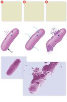
Phagocytosis
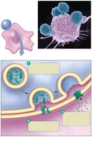
Phagocytic cells, such as neutrophils and macrophages, engulf and digest pathogens. The process involves capturing the bacterium, enclosing it in a vesicle, fusing with lysosomes, and digesting it.
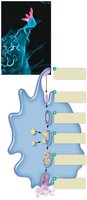
Inflammatory Response
Inflammation is triggered by histamine released from damaged cells and mast cells, causing blood vessels to dilate and become leaky. Complement proteins mark bacteria for destruction, and phagocytes attack pathogens.
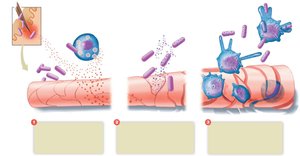
Specific Defense Mechanisms: The Third Line of Defense
The Immune Response
The immune response is highly specific, recognizing and targeting particular pathogens. It has memory and can distinguish between self and non-self cells.
Antigen: Any substance that triggers an immune response, usually a protein or polysaccharide on the surface of pathogens.
MHC Proteins: Major histocompatibility complex proteins are self-antigens that enable recognition of self cells.
Lymphocytes: B and T Cells
Lymphocytes are central to specific defenses. B lymphocytes mediate antibody immunity, while T lymphocytes mediate cell immunity.
B lymphocytes: Produce antibodies that neutralize antigens.
T lymphocytes: Directly attack foreign cells and coordinate immune responses.
B Cells: Antibody-Mediated Immunity
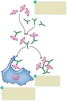
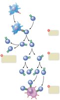
B cells are activated upon recognizing an antigen and divide into memory cells and plasma cells. Plasma cells secrete antibodies, while memory cells store information for future responses.
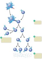
Antibody-Antigen Complex
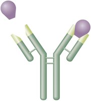
Antibodies bind to pathogens with matching surface antigens, forming antigen-antibody complexes. These complexes mark pathogens for attack by phagocytes or complement proteins.
Classes of Antibodies
Antibodies, also known as immunoglobulins (Ig), are classified into five types:
IgG: Most prevalent in blood.
IgM: First produced in immune response.
IgA: Found in body secretions.
IgD: Function unclear.
IgE: Key in allergic responses.
T Cells: Cell-Mediated Immunity
T cells originate from bone marrow stem cells and mature in the thymus. Types include CD4 (helper and memory T cells) and CD8 (cytotoxic T cells).
Helper T cells: Secrete cytokines to stimulate other immune cells.
Cytotoxic T cells: Attack and destroy abnormal or infected cells.
Memory T cells: Reactivate during later exposures.
Immune Memory
Immune memory allows for faster and stronger responses upon subsequent exposures to the same antigen.
Primary Response: Lag time of 3–6 days, peaks at 10–12 days.
Secondary Response: Lag time of hours, peaks in days, much more antibody produced.
Medical Assistance in Immunity
Immunization
Immunization is a strategy to develop immunity against specific pathogens. It can be active (vaccination) or passive (administering antibodies).
Monoclonal Antibodies
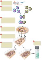
Monoclonal antibodies are produced in the laboratory by hybrid B cell clones and are used in diagnostic tests and treatments.
Antibiotics
Antibiotics combat bacteria by targeting features unique to bacterial cells. They are ineffective against viruses.
Tissue Rejection
Tissue rejection may occur after organ transplantation if the recipient’s immune system attacks the transplanted tissue. Matching ABO and MHC antigens and using immunosuppressive drugs minimize rejection risk.
Inappropriate Immune System Activity
Allergies
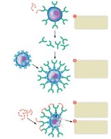
Allergies are hypersensitivity reactions to allergens, which are non-pathogenic substances. The response is mediated by IgE, basophils, mast cells, and histamine.
Localized: Affect only the exposed area.
Systemic: Affect multiple organ systems; anaphylactic shock is a severe systemic reaction.
Treatment: Antihistamines, epinephrine, allergy shots.
Autoimmune Disorders
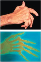
Autoimmune disorders occur when the immune system fails to distinguish self from non-self, attacking the body’s own tissues.
Lupus: Inflamed connective tissue, affecting various organs.
Rheumatoid Arthritis: Inflamed synovial membrane.
Immune Deficiency: AIDS
AIDS and HIV
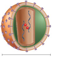
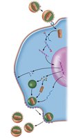
AIDS is caused by HIV, which targets helper T cells. HIV attaches to CD4 receptors and enters the cell. Transmission occurs via body fluids.
Progression of AIDS
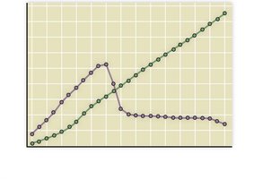
Phase I: Flu-like symptoms, virus multiplies, antibodies produced but ineffective.
Phase II: Opportunistic infections, decreasing helper T cells.
Phase III: Clinical AIDS, helper T cells below 200 cells/mm3, opportunistic infections and cancers.
The AIDS Epidemic
AIDS is a global health issue, with millions infected and dead worldwide. Most infections occur in sub-Saharan Africa, with increasing spread in Asia and India.
Risk Factors for AIDS
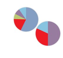
Risky behaviors increase the chance of acquiring AIDS. Males are more affected, primarily through sex with other men and intravenous drug use. Females are affected through sex with infected males and drug use.
Prevention and Treatment
Prevention: Abstinence, reducing sexual partners, using condoms, avoiding high-risk practices, and getting tested.
Treatment: Anti-HIV drugs inhibit entry, replication, integration, and assembly of viral proteins. Early treatment delays or prevents clinical AIDS. Vaccines are under development.
