 Back
The Cardiovascular System: Structure, Function, and Physiology
Study Guide - Smart Notes
 Tailored notes based on your materials, expanded with key definitions, examples, and context.
Cardiovascular System Overview
Blood Transport: Arteries, Capillaries & Veins
The cardiovascular system is a vast, branched network of hollow tubules responsible for transporting blood throughout the body. It consists of three major types of blood vessels: arteries, capillaries, and veins, each with distinct structural and functional characteristics.
Arteries: Thick-walled vessels that transport blood away from the heart. They are distensible, meaning they can stretch under pressure.
Capillaries: Microscopic vessels where exchange of solutes and water with cells occurs.
Veins: Thin-walled vessels that transport blood towards the heart.
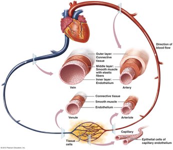
Structure & Function of Blood Vessels
Arteries
Arteries transport blood away from the heart to body tissues and are able to stretch due to blood pressure, storing blood from each heartbeat. Their structure is adapted to withstand high pressure:
Endothelium: Innermost layer of squamous cells, reduces friction.
Smooth Muscle: Thickest layer, elastic connective tissue for stretching and withstanding high pressure.
Connective Tissue: Outermost layer, primarily collagen, anchors vessels and protects from injury.
Aneurysm: A risk associated with high pressure in arteries, where blood seeps out of the endothelium and builds between outer layers, causing ballooning and potential rupture.
Arterioles
Arterioles are smaller branches of arteries with simpler structure, lacking connective tissue due to lower pressure. They regulate blood flow between arteries and capillaries using precapillary sphincters:
Vasoconstriction: Sphincters contract to reduce blood flow.
Vasodilation: Sphincters relax to increase blood flow.
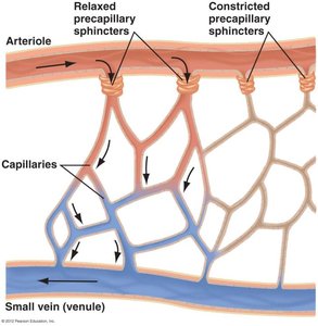
Capillaries: Blood Exchange
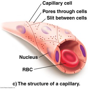
Capillaries connect arterioles and venules, forming an extensive network for exchange between blood and tissues. Their structure allows efficient diffusion:
Single layer of squamous cells (endothelium).
Very narrow diameter, causing RBCs to travel single file.
Pores and slits allow diffusion of nutrients, gases, and waste products.
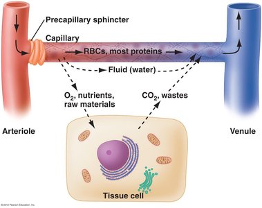
Fluid Movement in Capillaries
Fluid moves between capillaries, interstitial fluid, and cells through diffusion and filtration. Capillaries supply several nearby cells, facilitating exchange of oxygen, nutrients, and waste products.
Lymphatic System: Maintenance of Blood Volume
The lymphatic system returns excess plasma fluid, lipid droplets, and large substances from interstitial spaces back to the blood, maintaining blood volume. Lymphatic capillaries collect this fluid, which becomes lymph and is eventually returned to the bloodstream via veins near the heart.
Veins
Veins bring blood towards the heart, usually deoxygenated. Their structure is similar to arteries but with thinner walls and larger diameter. Veins accommodate large quantities of blood (about 2/3 of total blood volume) and operate under lower pressure.
Connective tissue: Thinner than in arteries.
Smooth muscle: Thinner than in arteries.
Epithelium: Same as arteries.
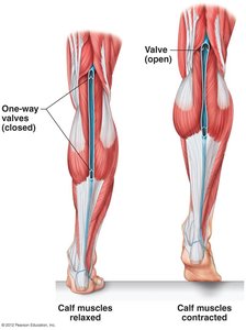
Veins have the ability to stretch and are assisted by mechanisms to return blood to the heart against gravity, such as the skeletal muscle pump and one-way valves.
Retrograde Flow of Blood
Three mechanisms assist veins in returning blood to the heart:
Skeletal muscles: Veins pass between muscles; contraction squeezes veins, pushing blood forward.
One-way valves: Prevent backflow, ensuring blood moves toward the heart.
Respiratory pump: Pressure changes during breathing push blood from abdomen to chest.
The Heart: Structure and Function
Heart Anatomy
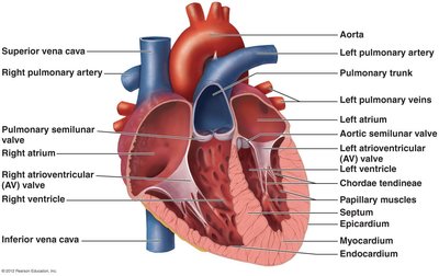
The heart is a muscular organ made of cardiac tissue, located in the thoracic cavity. It has four chambers:
Right and Left Atria: Receive blood.
Right and Left Ventricles: Pump blood out.
Septum: Muscular partition separating left and right sides.
The heart wall consists of three layers:
Epicardium: Outermost connective tissue layer.
Myocardium: Middle layer, responsible for contraction.
Endocardium: Innermost endothelial layer.
Heart Valves
Valves ensure unidirectional blood flow:
Atrioventricular (AV) valves: Between atria and ventricles (tricuspid and mitral/bicuspid).
Semilunar valves: Between ventricles and main arteries (aortic and pulmonary).
Chordae tendineae: Strong connective tissue strands supporting AV valves.
Circuits of Blood Flow
Pulmonary Circuit
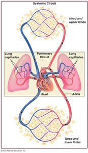
The pulmonary circuit carries deoxygenated blood from the right side of the heart to the lungs for gas exchange, then returns oxygenated blood to the left atrium.
Systemic Circuit
The systemic circuit carries oxygenated blood from the left ventricle through the aorta to the body, then returns deoxygenated blood to the right atrium via the vena cava.
Coronary Circulation
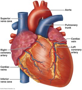
The heart muscle (myocardium) receives its own supply of oxygen and nutrients via the coronary arteries. Cardiac veins collect deoxygenated blood from the heart muscle and return it to the right atrium.
The Cardiac Cycle
Phases of the Cardiac Cycle
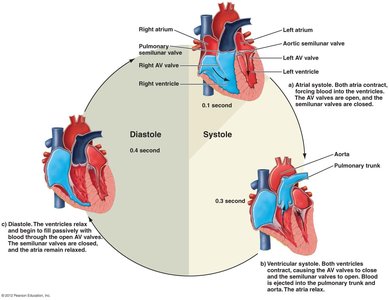
The cardiac cycle consists of contraction and relaxation phases:
Systole: Contraction phase (atria and ventricles contract).
Diastole: Relaxation phase (heart fills with blood).
The cycle takes about 0.8 seconds and includes:
Atrial systole (0.1 s): Atria contract, AV valves open, semilunar valves closed.
Ventricular systole (0.3 s): Ventricles contract, AV valves close, semilunar valves open.
Diastole (0.4 s): Both atria and ventricles relax, semilunar valves close, AV valves open.
Pulse and Heart Sounds
Pulse
The pulse is the expansion and recoil of arteries during systole and diastole, felt in arteries near the skin (e.g., carotid, radial, brachial).
Heart Sounds
"Lub": Closure of AV valves during ventricular systole.
"Dub": Closure of semilunar valves during ventricular diastole.
Heart murmurs: Unusual sounds due to incomplete valve closure.
Cardiac Conduction System
Electrical Flow in the Heart
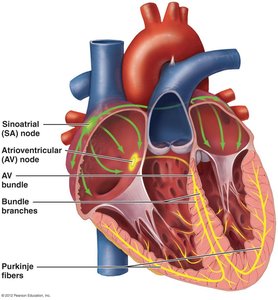
Specialized cardiac muscle cells initiate and distribute electrical impulses, coordinating the cardiac cycle. The conduction system includes:
Sinoatrial (SA) node: Pacemaker, initiates impulse.
Atrioventricular (AV) node: Delays impulse, allows atria to contract.
AV bundle: Conducts impulse to ventricles.
Purkinje fibers: Distribute impulse throughout ventricles.
Electrocardiogram (ECG/EKG)
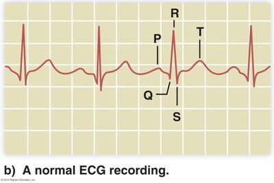
An ECG records electrical impulses in the heart. Key features:
P-wave: Impulse across atria.
QRS complex: Impulse through ventricles.
T-wave: End of ventricular electrical activity.
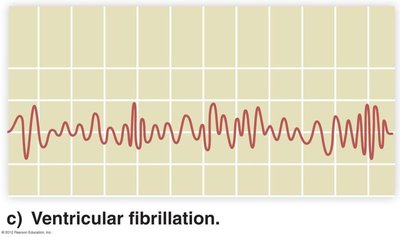
ECGs can detect irregularities such as arrhythmia and ventricular fibrillation.
Blood Pressure
Definition and Components
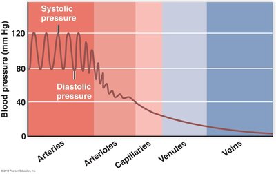
Blood pressure is the force exerted by blood on vessel walls due to heart pumping. It varies across vessel types:
Systolic pressure: Highest, during ventricular contraction.
Diastolic pressure: Lowest, during ventricular relaxation.
Blood Pressure Categories
Blood Pressure Category | Systolic (mm Hg) | Diastolic (mm Hg) |
|---|---|---|
Normal | Less than 120 | Less than 80 |
Elevated | 120-129 | Less than 80 |
Hypertension Stage 1 | 130-139 | 80-89 |
Hypertension Stage 2 | ≥140 | ≥90 |
Hypertensive Crisis | Greater than 180 | Greater than 120 |
Key Equations
Blood Pressure Equation
Blood pressure is often measured as:
Cardiac Output Equation
Cardiac output is the volume of blood pumped by the heart per minute:
Summary
The cardiovascular system is essential for transporting blood, nutrients, gases, and waste products throughout the body. Its structure and function are tightly regulated to maintain homeostasis and support cellular activity.
