 Back
The Cardiovascular System: Structure, Function, and Regulation
Study Guide - Smart Notes
 Tailored notes based on your materials, expanded with key definitions, examples, and context.
Cardiovascular System Overview
Blood Transport: Arteries, Capillaries, and Veins
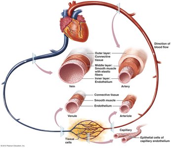
The cardiovascular system is a vast network responsible for transporting blood throughout the body. It consists of three major types of blood vessels: arteries, capillaries, and veins, each with distinct structural and functional characteristics.
Arteries: Thick-walled vessels that transport blood away from the heart. Their elasticity allows them to stretch under pressure and recoil, maintaining blood flow between heartbeats.
Capillaries: Microscopic vessels where exchange of solutes, gases, and water occurs between blood and tissues. Their thin walls facilitate efficient diffusion.
Veins: Thin-walled vessels that return blood toward the heart. They accommodate large volumes of blood and rely on various mechanisms to overcome gravity.
Additional info: The lumen is the hollow interior of blood vessels, crucial for blood flow.
Structure and Function of Blood Vessels
Arteries and Arterioles
Arteries are composed of three layers:
Endothelium: Innermost layer of squamous cells, reduces friction.
Smooth Muscle: Middle layer, thickest, contains elastic fibers for stretching and recoil.
Connective Tissue: Outermost layer, primarily collagen, anchors and protects vessels.
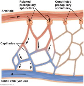
Arterioles are smaller branches of arteries, lacking connective tissue due to lower pressure. They regulate blood flow into capillaries via precapillary sphincters, which contract (vasoconstriction) or relax (vasodilation) to control blood entry.
Capillaries: Sites of Exchange
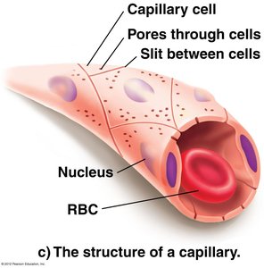
Capillaries connect arterioles and venules, forming an extensive network. Their walls consist of a single layer of squamous cells, allowing for efficient exchange of gases, nutrients, and waste products. Red blood cells (RBCs) travel single file due to the narrow diameter.
Diffusion: Nutrients and waste products move through pores and slits in capillary walls.
Gas Exchange: Oxygen and carbon dioxide easily diffuse across capillary membranes.
Fluid Movement and Exchange
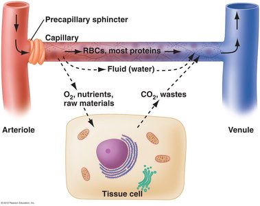
Fluid moves between capillaries, interstitial fluid, and cells through a combination of hydrostatic and osmotic pressures. Precapillary sphincters regulate the flow, ensuring that each cell receives adequate nutrients and oxygen.
Osmosis and Diffusion: Water, oxygen, nutrients, and waste products are exchanged between blood and tissue cells.
Role of Sphincters: Control which capillaries are open for exchange.
Lymphatic System: Maintenance of Blood Volume
Role in Fluid Balance
The lymphatic system returns excess plasma fluid, lipid droplets, and large substances from interstitial spaces back to the bloodstream, maintaining blood volume. Lymphatic capillaries collect this fluid, which becomes lymph and is eventually returned to veins near the heart.
Excess Fluid: Approximately 2-3 liters per day are returned via the lymphatic system.
Composition: Lymph does not contain plasma proteins or RBCs.
Veins: Blood Return Mechanisms
Structure and Function
Veins have thinner walls and larger diameters than arteries, with less smooth muscle and connective tissue. They stretch to accommodate large volumes of blood, and most blood in the body is found in veins.
Varicose Veins: Result from prolonged standing, causing permanent swelling.
Blood Return: Veins rely on three mechanisms to return blood to the heart:
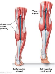
Skeletal Muscle Pump: Muscles contract and squeeze veins, pushing blood toward the heart.
One-way Valves: Prevent backflow, ensuring unidirectional movement.
Respiratory Pump: Pressure changes during breathing move blood from abdomen to chest.
The Heart: Structure and Function
Anatomy of the Heart
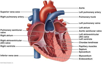
The heart is a muscular organ located in the thoracic cavity. It consists of four chambers: two atria and two ventricles, separated by a septum. The heart wall has three layers: epicardium (outer), myocardium (middle, responsible for contraction), and endocardium (inner).
Pericardium: Fibrous outer layer that protects and anchors the heart.
Pericardial Cavity: Contains lubricating fluid for smooth contractions.
Heart Valves
Valves ensure unidirectional blood flow:
Atrioventricular (AV) Valves: Tricuspid (right) and mitral/bicuspid (left) valves control flow between atria and ventricles.
Semilunar Valves: Prevent backflow from arteries into ventricles; include aortic and pulmonary valves.
Chordae Tendineae: Strong connective tissue strands supporting AV valves.
Circuits of Blood Flow
Pulmonary and Systemic Circuits
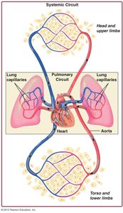
The heart pumps blood through two main circuits:
Pulmonary Circuit: Right side of heart sends deoxygenated blood to lungs for gas exchange.
Systemic Circuit: Left side of heart sends oxygenated blood to the body.
Coronary Circulation
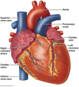
The myocardium receives oxygen and nutrients via coronary arteries. Cardiac veins collect deoxygenated blood and return it to the right atrium.
The Cardiac Cycle
Phases of the Cardiac Cycle
The cardiac cycle consists of alternating contraction (systole) and relaxation (diastole) phases:
Atrial Systole (0.1 s): Atria contract, filling ventricles.
Ventricular Systole (0.3 s): Ventricles contract, ejecting blood.
Diastole (0.4 s): Heart relaxes, chambers refill with blood.
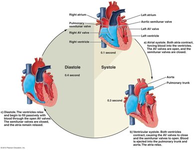
Additional info: The "lub" sound is from AV valve closure; "DUB" is from semilunar valve closure.
Cardiac Conduction System
Electrical Flow in the Heart
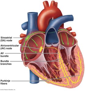
Specialized cardiac muscle cells coordinate the heartbeat:
Sinoatrial (SA) Node: Initiates electrical impulse; acts as pacemaker.
Atrioventricular (AV) Node: Delays impulse, allowing atria to contract.
AV Bundle and Purkinje Fibers: Distribute impulse to ventricles, causing coordinated contraction.
Electrocardiogram (ECG/EKG)
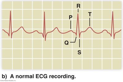
An ECG records the electrical activity of the heart. Key components:
P-wave: Atrial depolarization
QRS complex: Ventricular depolarization
T-wave: Ventricular repolarization
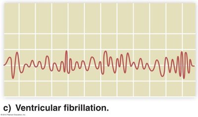
ECGs can detect arrhythmias and ventricular fibrillation, which are abnormal heart rhythms.
Blood Pressure
Definition and Components
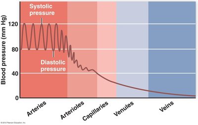
Blood pressure is the force exerted by blood on vessel walls, resulting from heart contractions. It varies across vessel types:
Systolic Pressure: Highest during ventricular contraction.
Diastolic Pressure: Lowest during ventricular relaxation.
Normal BP: 120/80 mm Hg or lower.
Blood Pressure Categories
Blood pressure is classified as follows:
Category | Systolic (mm Hg) | Diastolic (mm Hg) |
|---|---|---|
Normal | Less than 120 | Less than 80 |
Elevated | 120-129 | Less than 80 |
Hypertension Stage 1 | 130-139 | 80-89 |
Hypertension Stage 2 | ≥140 | ≥90 |
Hypertensive Crisis | Greater than 180 | Greater than 120 |
Additional info: Persistent high blood pressure (hypertension) increases risk for heart disease and stroke.
