 Back
The Heart and Blood Vessels: Structure, Function, and Regulation
Study Guide - Smart Notes
 Tailored notes based on your materials, expanded with key definitions, examples, and context.
The Heart and Blood Vessels
Blood Vessels Transport Blood
The cardiovascular system is composed of a network of blood vessels that transport blood throughout the body. These vessels include arteries, arterioles, capillaries, venules, and veins, each with distinct structural and functional properties.
Arteries: Transport blood away from the heart. Large arteries have thick muscular walls, are stiff yet elastic, and their elastic recoil maintains blood pressure between heartbeats.
Endothelium: The thin inner layer of blood vessels, composed of flat squamous epithelial cells, creates a slick surface to minimize friction and promote smooth blood flow.
Muscle and Connective Tissue Layers: Outside the endothelium, a thick layer of smooth muscle and elastic connective tissue resists internal pressure and allows for elastic recoil. The outermost layer consists of tough connective tissue (mostly collagen) for protection.
Aneurysm: Ballooning of the artery wall, which can bulge inward or outward, potentially causing obstruction, chest pain, or rupture.
Arterioles and Precapillary Sphincters
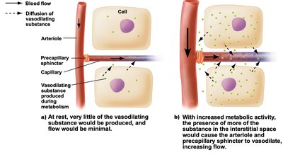
Arterioles are smaller arteries that regulate blood flow to capillaries by contracting or relaxing their smooth muscle layer. Precapillary sphincters, bands of smooth muscle at the junction of arterioles and capillaries, act as gates to control blood flow.
Vasodilation: Relaxation of smooth muscle increases blood flow to capillaries.
Vasoconstriction: Contraction reduces blood flow.
Regulation is influenced by nerves, hormones, and local environmental conditions (e.g., temperature).
Capillaries
Capillaries are thin-walled, narrow vessels forming extensive networks (capillary beds) that facilitate exchange of gases, nutrients, and waste products between blood and tissues.
Single layer of squamous epithelial cells with narrow slits and tiny pores.
Allow exchange of fluids, while retaining red blood cells and plasma proteins; some white blood cells can squeeze through.
Exchange of interstitial fluid occurs only in capillaries.
Most filtered fluid returns to the blood; excess is managed by the lymphatic system.
Lymphatic System
The lymphatic system absorbs excess plasma fluid and returns it to the venous system, playing a role in immune defense and maintaining proper blood and interstitial fluid volumes.
Similar to the venous system but lacks plasma proteins and red blood cells.
Transports lymph back to veins near the heart.
Not part of the cardiovascular system.
Veins
Veins return blood to the heart and serve as reservoirs for blood volume. Their walls are thinner and more distensible than arteries, allowing them to accommodate large volumes at low pressure.
Contain one-way valves to prevent backflow.
Three mechanisms assist venous return: skeletal muscle pump, valves, and respiratory pump.
Problems such as varicose veins can arise from blood pooling due to gravity.
The Heart Pumps Blood Through the Vessels
Structure of the Heart
The heart is a muscular organ enclosed by the pericardium, which protects and anchors it. The heart wall consists of three layers: epicardium (outer), myocardium (middle, muscular), and endocardium (inner).
Epicardium: Thin outer layer of connective and epithelial tissue.
Myocardium: Thick middle layer of cardiac muscle responsible for contraction.
Endocardium: Thin inner layer continuous with blood vessel endothelium.
Heart Chambers and Valves
The heart contains four chambers: two atria (upper) and two ventricles (lower), separated by the septum. Four one-way valves prevent backflow of blood.
Right atrium and ventricle: Pump blood to the lungs.
Left atrium and ventricle: Pump blood to the rest of the body.
Atrioventricular (AV) valves: Tricuspid (right, three flaps) and bicuspid/mitral (left, two flaps).
Semilunar valves: Prevent backflow from arteries into ventricles.
Pattern of Blood Flow
The heart pumps blood through two circuits: pulmonary (lungs) and systemic (body). Blood passes through the heart twice per cycle—once as deoxygenated blood for the lungs, then as oxygenated blood for the body.
Coronary arteries supply the heart muscle; cardiac veins return blood to the right atrium.
The Cardiac Cycle
The cardiac cycle consists of systole (contraction) and diastole (relaxation). Each cycle involves coordinated contraction and relaxation of atria and ventricles, regulated by the cardiac conduction system.
Atrial systole: Atria contract, filling ventricles.
Ventricular systole: Ventricles contract, AV valves close, semilunar valves open.
Diastole: Both atria and ventricles relax; semilunar valves close, AV valves open.
Heart sounds: Caused by valve closures; murmurs indicate abnormal flow.
Cardiac Conduction System
The cardiac conduction system initiates and distributes electrical impulses, coordinating heart contractions. Key components include the sinoatrial (SA) node, atrioventricular (AV) node, AV bundle, bundle branches, and Purkinje fibers.
SA node: Cardiac pacemaker; initiates electrical signals.
AV node: Delays impulse, allowing atria to contract before ventricles.
AV bundle and Purkinje fibers: Distribute impulse to ventricles.
Electrocardiogram (ECG)
An ECG records the heart's electrical activity, tracking P waves (atrial impulse), QRS complex (ventricular impulse), and T waves (ventricular recovery). Arrhythmias are abnormal rhythms; ventricular fibrillation is life-threatening and treated by cardioversion.
Recap
The heart wall consists of three layers: the epicardium, the myocardium, and the endocardium. The heart contains four chambers and four one-way valves. The right atrium and right ventricle pump blood to the lungs; the left atrium and left ventricle pump blood to the rest of the body. Each cardiac cycle is a repetitive sequence of contraction (systole) and relaxation (diastole). Contraction of the heart is coordinated by modified cardiac muscle cells that initiate and transmit electrical impulses through a specialized conduction system. An electrocardiogram (ECG) is a recording of the heart’s electrical activity taken from the surface of the body.
Blood Pressure and Regulation
Blood Pressure
Blood pressure is the force exerted by blood on vessel walls, resulting from heart pumping. It varies across vessels and is measured in mm Hg using a sphygmomanometer.
Systolic pressure: Pressure during ventricular contraction; healthy value < 120 mm Hg.
Diastolic pressure: Pressure during relaxation; desirable value < 80 mm Hg.
Hypertension
High blood pressure increases risk for cardiovascular disease, heart attack, stroke, and kidney damage. Often caused by vessel narrowing (atherosclerosis). True hypertension is sustained elevation; prehypertension and isolated systolic hypertension are variations.
Hypotension
Blood pressure that is too low can also be problematic.
Regulation of the Cardiovascular System
Homeostatic regulation centers on maintaining constant arterial blood pressure by adjusting cardiac output, arteriole diameter, and blood volume.
Baroreceptors: Pressure sensors in the aorta and carotid arteries monitor blood pressure and signal the brain to adjust heart rate and vessel diameter.
Cardiac output: Amount of blood pumped per minute; calculated as heart rate × stroke volume.
Resting cardiac output: liters/minute
Medulla oblongata: Cardiovascular center receives inputs and sends outputs via sympathetic (raises BP) and parasympathetic (lowers BP) nerves.
Epinephrine and norepinephrine: Hormones increase cardiac output during sympathetic activation.
Local Blood Flow Regulation
Precapillary sphincters fine-tune blood flow to tissues based on metabolic activity. Vasodilation increases flow to active tissues; vasoconstriction shunts blood to vital organs during low pressure.
Cardiovascular Disorders
Common Disorders
Angina: Pain and tightness in the chest due to narrowed coronary arteries; treated with medication, angioplasty, or bypass graft.
Heart Attack: Sudden death of myocardium; diagnosed by ECG and blood enzymes; treated with arrhythmia control, clot-dissolving medications, or bypass graft.
Heart Failure: Weak heart muscle leads to fluid backup; treatment prevents fluid accumulation and improves cardiac performance.
Embolism: Sudden blockage by floating material (cholesterol, tissue, cancer); can cause pulmonary embolism, stroke, or heart attack.
Stroke: Brain damage from lack of blood, caused by embolism or artery rupture.
Heart Replacement
Heart transplants and artificial hearts are used for severe heart failure; xenotransplants (from animals) are a potential solution for organ shortages.
Disorder | Main Cause | Symptoms | Treatment |
|---|---|---|---|
Angina | Narrowed coronary arteries | Chest pain, tightness | Medication, angioplasty, bypass graft |
Heart Attack | Blocked coronary artery | Intense chest pain, nausea | Arrhythmia control, clot-dissolving meds, bypass graft |
Heart Failure | Weak heart muscle | Fluid backup, fatigue | Prevent fluid accumulation, improve performance |
Embolism | Floating material in blood | Chest pain, stroke, heart attack | Depends on location and cause |
Stroke | Embolism or artery rupture | Brain damage, loss of function | Emergency intervention |
Additional info: The notes have been expanded to include definitions, examples, and a summary table for cardiovascular disorders, as well as detailed explanations of blood vessel structure and function, cardiac cycle, and regulation mechanisms.
