 Back
The Heart and Blood Vessels: Structure, Function, and Regulation
Study Guide - Smart Notes
 Tailored notes based on your materials, expanded with key definitions, examples, and context.
The Heart and Blood Vessels
Overview
The cardiovascular system is responsible for transporting blood throughout the body, delivering oxygen and nutrients, and removing waste products. It consists of the heart, blood vessels (arteries, arterioles, capillaries, venules, and veins), and is closely associated with the lymphatic system.
Blood Vessels: Structure and Function
Arteries
Arteries transport blood away from the heart under high pressure.
Large arteries have thick layers of smooth muscle and elastic connective tissue, allowing them to stretch and recoil, maintaining blood pressure between heartbeats.
The endothelium is the thin, inner layer of flat squamous epithelial cells, providing a smooth, low-friction surface for blood flow.
The outermost layer is tough connective tissue (mainly collagen), protecting arteries from injury.
Aneurysm: A ballooning of the artery wall, which can lead to rupture or impaired blood flow.
Arterioles and Precapillary Sphincters
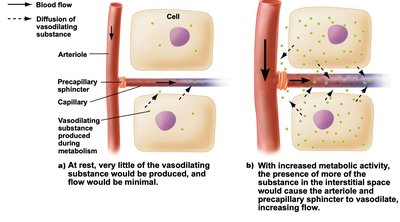
Arterioles are smaller branches of arteries with thinner walls, primarily regulating blood flow to capillaries by contracting or relaxing their smooth muscle layer.
At the junction with capillaries, precapillary sphincters (bands of smooth muscle) act as gates to control blood flow into capillary beds.
Vasodilation (relaxation) increases blood flow; vasoconstriction (contraction) decreases it. These processes are influenced by nerves, hormones, and local environmental conditions (e.g., temperature).
Capillaries
Capillaries are the smallest, thinnest blood vessels, forming extensive networks called capillary beds.
They consist of a single layer of squamous epithelial cells with narrow slits and pores, allowing exchange of gases, nutrients, and waste between blood and tissues.
Red blood cells and plasma proteins generally remain inside, while some white blood cells can pass through.
Most filtered fluid is reabsorbed into capillaries; excess interstitial fluid is collected by the lymphatic system.
Lymphatic System
Collects excess interstitial fluid and large substances (e.g., lipid droplets) not reabsorbed by capillaries.
Returns lymph to the venous system near the heart and plays a role in immune defense.
Helps maintain proper blood and interstitial fluid volume.
Veins and Venules
Veins return blood to the heart at lower pressure and have thinner walls and larger diameters than arteries.
Veins serve as blood reservoirs, containing about two-thirds of the body's blood volume.
Mechanisms assisting venous return include:
Skeletal muscle pump: Muscle contractions squeeze veins, pushing blood toward the heart.
One-way valves: Prevent backflow of blood.
Respiratory pump: Pressure changes during breathing move blood from abdominal to thoracic veins.
Varicose veins can develop if blood pools in the legs due to valve failure.
The Heart: Structure and Function
Heart Wall and Chambers
The heart wall consists of three layers:
Epicardium: Outer, thin layer of connective and epithelial tissue.
Myocardium: Thick, middle layer of cardiac muscle responsible for contraction.
Endocardium: Inner, thin layer of connective tissue, continuous with blood vessel endothelium.
The heart contains four chambers: two atria (upper) and two ventricles (lower), separated by a muscular septum.
Four one-way valves ensure unidirectional blood flow:
Atrioventricular (AV) valves: Tricuspid (right) and bicuspid/mitral (left), supported by chordae tendineae and papillary muscles.
Semilunar valves: Pulmonary and aortic, prevent backflow into ventricles.
Blood Flow Through the Heart
The heart pumps blood through two circuits simultaneously:
Pulmonary circuit: Right atrium → right ventricle → pulmonary arteries → lungs (gas exchange) → pulmonary veins → left atrium.
Systemic circuit: Left atrium → left ventricle → aorta → body tissues → veins → right atrium.
The left ventricle is the most muscular chamber, generating high pressure to pump blood throughout the body.
The heart's own blood supply comes from the coronary arteries; blood is collected by cardiac veins and returned to the right atrium.
The Cardiac Cycle
The cardiac cycle consists of:
Systole: Contraction phase (atria then ventricles).
Diastole: Relaxation phase (atria and ventricles fill with blood).
Heart sounds are produced by valve closures:
First sound: AV valves closing during ventricular systole.
Second sound: Semilunar valves closing during ventricular diastole.
Murmurs may indicate valve problems.
Cardiac Conduction System
Coordinated contraction is controlled by the cardiac conduction system:
Sinoatrial (SA) node: Pacemaker, initiates electrical impulses.
Atrioventricular (AV) node: Delays impulse, allowing atria to contract before ventricles.
Atrioventricular bundle and Purkinje fibers: Distribute impulse through ventricles.
An electrocardiogram (ECG) records the heart's electrical activity, showing P waves (atria), QRS complex (ventricles), and T waves (ventricular recovery).
Arrhythmias are irregular heart rhythms; severe forms like ventricular fibrillation require immediate treatment (e.g., cardioversion, CPR).
Blood Pressure and Its Regulation
Blood Pressure
Blood pressure is the force exerted by blood on vessel walls, measured in mm Hg.
Normal values: Systolic < 120 mm Hg, Diastolic < 80 mm Hg.
Measured using a sphygmomanometer.
Hypertension and Hypotension
Hypertension: Sustained high blood pressure, a major risk factor for cardiovascular disease ("silent killer").
Can lead to heart attack, stroke, kidney damage; often caused by narrowed arteries (atherosclerosis).
Hypotension: Abnormally low blood pressure.
Regulation of the Cardiovascular System
Homeostasis centers on maintaining constant arterial blood pressure by regulating:
Cardiac output (heart rate × stroke volume)
Arteriole diameter
Blood volume
Baroreceptors in the aorta and carotid arteries detect pressure changes and signal the brain to adjust heart rate, force of contraction, and vessel diameter.
The medulla oblongata (cardiovascular center) integrates these signals, using sympathetic (increase BP) and parasympathetic (decrease BP) nerves, as well as hormones (epinephrine, norepinephrine).
Local blood flow is fine-tuned by precapillary sphincters in response to tissue needs.
Cardiovascular Disorders
Common Disorders
Angina: Chest pain due to reduced blood flow in coronary arteries; diagnosed with angiography, treated with medication, angioplasty, or bypass surgery.
Heart Attack (Myocardial Infarction): Death of heart muscle tissue due to blocked blood supply; symptoms include chest pain, nausea; diagnosed by ECG and blood tests.
Heart Failure: Weakness of heart muscle, leading to fluid buildup (congestive heart failure); managed by medications and lifestyle changes.
Embolism: Sudden blockage of a blood vessel by a floating object (e.g., clot, fat, air); can cause pulmonary embolism, stroke, or heart attack.
Stroke: Brain damage from interrupted blood supply, often due to embolism or artery rupture.
Treatments and Interventions
Heart transplant for end-stage heart failure.
Artificial hearts and xenotransplants (from animals) as temporary or experimental solutions.
Recap
The heart wall consists of three layers: the epicardium, the myocardium, and the endocardium. The heart contains four chambers and four one-way valves. The right atrium and right ventricle pump blood to the lungs; the left atrium and left ventricle pump blood to the rest of the body. Each cardiac cycle is a repetitive sequence of contraction (systole) and relaxation (diastole). Contraction of the heart is coordinated by modified cardiac muscle cells that initiate and transmit electrical impulses through a specialized conduction system. An electrocardiogram (ECG) is a recording of the heart’s electrical activity taken from the surface of the body.
