 Back
The Human Respiratory System: Structure, Function, and Gas Exchange
Study Guide - Smart Notes
 Tailored notes based on your materials, expanded with key definitions, examples, and context.
Respiratory System: Exchange of Gases
Overview of Human Respiratory System
The human respiratory system is responsible for the intake of oxygen and the removal of carbon dioxide, facilitating gas exchange essential for cellular respiration. It is divided into upper and lower respiratory tracts, each with specialized structures and functions.
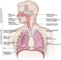
Respiration Processes
Respiration involves several interconnected processes that ensure oxygen delivery and carbon dioxide removal:
Breathing (Ventilation): The physical movement of air into and out of the lungs.
External Respiration: Gas exchange between air in the alveoli and blood in pulmonary capillaries.
Internal Respiration: Gas exchange between blood and tissue cells throughout the body.
Cellular Respiration: The use of oxygen by cells to produce ATP, with carbon dioxide as a waste product.
Structure and Function of the Respiratory Tract
Components of the Upper Respiratory Tract
The upper respiratory tract includes the nasal cavity, nose, sinuses, and pharynx. These structures filter, warm, and moisten incoming air, and serve as passageways for respiration and olfaction.
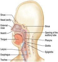
Functions of the Upper Respiratory Tract
Passageway for Respiration: Conducts air from the external environment to the lower respiratory tract.
Receptors for Smell: Located in the superior nasal cavity.
Filtration: Removes larger foreign particles from incoming air.
Moistening and Warming: Prepares air for entry into the lungs.
Resonating Chambers: Sinuses contribute to voice resonance.
Components of the Lower Respiratory Tract
The lower respiratory tract consists of the larynx, trachea, bronchi, bronchioles, and alveoli. These structures facilitate the transport of air and the exchange of gases.
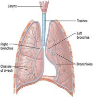
Functions of the Lower Respiratory Tract
Larynx: Maintains an open airway, routes food and air, and assists in sound production.
Trachea: Transports air to and from the lungs.
Bronchi: Branch into the lungs, directing air to each lung.
Lungs: Transport air to alveoli for gas exchange.
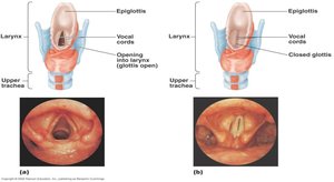
Anatomy of the Larynx
The larynx, or voice box, is a critical structure for maintaining an open airway and producing sound. It contains the vocal cords and the epiglottis, which prevents food from entering the airway during swallowing.
Anatomy of the Trachea and Cough Reflex
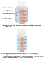
The trachea is composed of incomplete rings of hyaline cartilage joined by smooth muscle. During coughing, the smooth muscle contracts, decreasing the diameter of the trachea and increasing the force of air expulsion.
Pleural Membranes
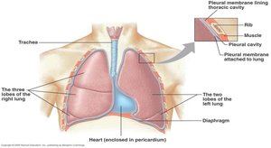
Pleural membranes surround the lungs and line the thoracic cavity, creating a pleural cavity filled with fluid. This arrangement reduces friction during breathing and helps maintain lung expansion.
Gas Exchange and Transport
Gas Exchange Between the Blood and Alveoli
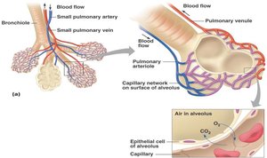
Gas exchange occurs in the alveoli, where oxygen diffuses into the blood and carbon dioxide diffuses out. This process is driven by differences in partial pressures of gases.
Respiratory Cycle: Inspiration and Expiration
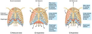
The respiratory cycle consists of inspiration and expiration, regulated by changes in thoracic volume and pressure. The diaphragm and intercostal muscles play key roles in this process.
Relaxed State: Diaphragm and intercostal muscles are relaxed.
Inspiration: Diaphragm contracts and moves downward, intercostal muscles elevate the chest wall, increasing thoracic volume and decreasing pressure, drawing air in.
Expiration: Muscles relax, diaphragm resumes dome shape, chest lowers, increasing pressure and expelling air.
Measurement of Lung Function
Spirometry and Lung Volumes
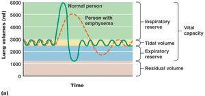
Spirometry is used to measure lung volumes and capacities, providing important information about respiratory health.
Tidal Volume: Volume of air inhaled and exhaled in a single breath.
Dead Space Volume: Air remaining in airways not participating in gas exchange.
Vital Capacity: Maximal volume exhaled after maximal inhalation.
Inspiratory Reserve Volume: Additional air inhaled beyond tidal volume.
Expiratory Reserve Volume: Additional air exhaled beyond tidal volume.
Residual Volume: Air remaining in lungs after forceful expiration.
Gas Exchange and Transport Mechanisms
Pulmonary and Systemic Circuits
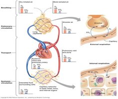
The pulmonary circuit carries blood between the heart and lungs for gas exchange, while the systemic circuit distributes oxygenated blood to tissues and returns deoxygenated blood to the heart.
How Oxygen and Carbon Dioxide are Carried
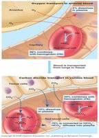
Oxygen is primarily transported bound to hemoglobin in red blood cells, while carbon dioxide is carried dissolved in plasma, bound to hemoglobin, or as bicarbonate ions.
Oxygen Transport: 98% bound to hemoglobin, 2% dissolved in plasma.
Carbon Dioxide Transport: 10% dissolved in plasma, 20% bound to hemoglobin, 70% as plasma bicarbonate.
Gas Exchange and Transport: Passive Diffusion
Gases diffuse according to their partial pressures. External respiration exchanges gases between air and blood, while internal respiration exchanges gases with tissue fluids.
Gas | Transport Mechanism |
|---|---|
Oxygen | Hemoglobin (98%), Plasma (2%) |
Carbon Dioxide | Plasma (10%), Hemoglobin (20%), Bicarbonate (70%) |
Regulation of Breathing
Nervous System Involvement
The respiratory center in the medulla oblongata establishes the basic breathing pattern. Chemical receptors monitor levels of carbon dioxide, hydrogen ions, and oxygen, adjusting breathing rate as needed.
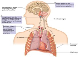
Medulla Oblongata: Sensitive to hydrogen ions in cerebrospinal fluid, which result from carbon dioxide in blood.
Carotid and Aortic Bodies: Sensitive to carbon dioxide, pH, and oxygen levels.
Conscious Control: Higher brain centers can modify breathing voluntarily.
Disorders of the Respiratory System
Common Disorders
The respiratory system can be affected by various disorders, impacting airflow, gas exchange, and overall health.
Reduced Air Flow: Asthma, emphysema, bronchitis
Infections: Pneumonia, tuberculosis, botulism
Lung Cancer
Congestive Heart Failure
Cystic Fibrosis
Summary Table: Respiratory System Disorders
Disorder | Main Effect |
|---|---|
Asthma | Reduced airflow due to bronchial constriction |
Emphysema | Destruction of alveoli, reduced gas exchange |
Bronchitis | Inflammation of bronchi, increased mucus |
Pneumonia | Infection of alveoli, impaired gas exchange |
Tuberculosis | Bacterial infection, lung tissue damage |
Botulism | Paralysis of respiratory muscles |
Lung Cancer | Uncontrolled cell growth, impaired function |
Congestive Heart Failure | Fluid accumulation in lungs |
Cystic Fibrosis | Thick mucus, blocked airways |
Additional info: The notes expand on brief points with academic context, definitions, and examples to ensure completeness and clarity for exam preparation.
