 Back
The Muscular System: Structure, Function, and Contraction
Study Guide - Smart Notes
 Tailored notes based on your materials, expanded with key definitions, examples, and context.
L The Muscular System
Introduction to Muscles and Muscle Tissue
Muscle tissue is specialized for contraction, enabling movement of the body and its parts. Muscles convert chemical energy (from ATP) into mechanical energy, generating force and movement. The main properties of muscle tissue are essential for their function in the human body.
Contractility: The ability of muscle tissue to shorten forcibly when stimulated, producing movement or tension.
Extensibility: The capacity of muscle cells to be stretched or extended without being damaged.
Elasticity: The ability of muscle tissue to return to its original length after being stretched or contracted.
Excitability: The ability to respond to stimuli, usually from the nervous system.
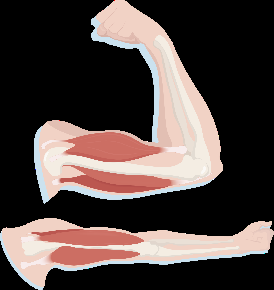
Types of Muscle Tissue
There are three main types of muscle tissue in the human body, each with distinct structure and function:
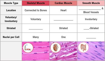
Skeletal Muscle: Attached to bones, responsible for voluntary movements. Cells are long, multinucleated, and striated.
Cardiac Muscle: Found only in the heart, responsible for pumping blood. Cells are branched, striated, and typically have one nucleus. Contraction is involuntary.
Smooth Muscle: Located in the walls of hollow organs (e.g., intestines, blood vessels). Cells are spindle-shaped, non-striated, and have a single nucleus. Contraction is involuntary.
Muscle Type | Location | Voluntary/Involuntary | Striated | Nuclei per Cell |
|---|---|---|---|---|
Skeletal Muscle | Connected to Bones | Voluntary | Striated | Many |
Cardiac Muscle | Heart | Involuntary | Striated | One |
Smooth Muscle | Blood Vessels & Organs | Involuntary | Non-Striated | One |
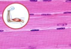
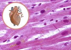
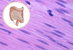
Structure of a Skeletal Muscle
Organization of Muscle Tissue
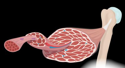
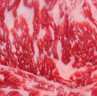
Skeletal muscles are complex organs composed of muscle fibers, nerves, blood vessels, and connective tissue. The organization of these components allows for efficient force generation and transmission.
Muscle Fiber: A single muscle cell, long and multinucleated, surrounded by endomysium.
Fascicle: A bundle of muscle fibers, surrounded by perimysium.
Muscle: A bundle of fascicles, surrounded by epimysium.
Connective Tissue: Connects muscle to bone via tendons (cord-like) or aponeuroses (sheet-like).

The Muscle Fiber
Each muscle fiber contains specialized structures for contraction:
Sarcolemma: The plasma membrane of a muscle fiber, wrapping bundles of myofibrils.
Myofibrils: Long, rod-shaped organelles containing myofilaments (actin and myosin).
Sarcoplasmic Reticulum: Specialized endoplasmic reticulum that stores and releases calcium ions (Ca2+).
T-Tubules: Invaginations of the sarcolemma that conduct action potentials deep into the muscle fiber.
Hierarchy of Muscle Structure
Superficial to Deep: Epimysium → Perimysium → Fascicle → Endomysium → Muscle Fiber → Sarcolemma → Myofibrils → Myofilaments
Sliding Filament Theory and the Sarcomere
Structure of the Sarcomere
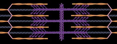
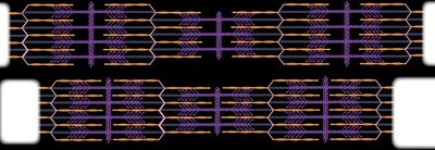
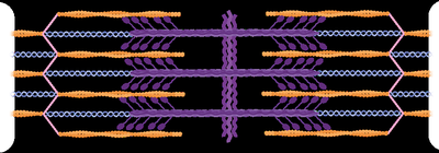
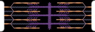
The sarcomere is the fundamental contractile unit of skeletal muscle, defined by the arrangement of actin (thin) and myosin (thick) filaments. The interaction between these filaments produces muscle contraction.
Myosin: Thick filament, anchored at the M line (center of sarcomere).
Actin: Thin filament, anchored at the Z disc (ends of sarcomere).
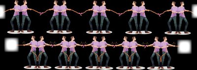
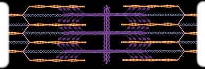
During contraction, actin filaments slide toward the center, increasing overlap with myosin; the sarcomere shortens, but the filaments themselves do not change length.

Proteins of the Sarcomere
Contractile Proteins: Myosin (thick), Actin (thin)
Regulatory Proteins: Tropomyosin (blocks myosin binding sites on actin), Troponin (binds Ca2+, moves tropomyosin to expose binding sites)
Structural Proteins: Titin (elastic filament, maintains sarcomere structure)


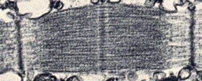
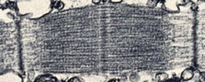
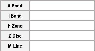
Sarcomere Bands, Zones, Discs, and Lines
Z Disc: End of the sarcomere, anchors actin filaments.
M Line: Center of the sarcomere, anchors myosin filaments.
I Band: Region with only actin (thin filaments).
A Band: Region with both actin and myosin overlap.
H Zone: Center region with only myosin (thick filaments).
Region | Description |
|---|---|
A Band | Area with both actin and myosin filaments |
I Band | Area with only actin filaments |
H Zone | Area with only myosin filaments |
Z Disc | Boundary of the sarcomere, anchors actin |
M Line | Center of the sarcomere, anchors myosin |
Steps of Muscle Contraction
Excitation-Contraction Coupling
Muscle contraction is initiated by a nerve impulse (action potential) that travels to the muscle fiber, leading to the release of calcium ions and the exposure of myosin binding sites on actin.
Action potential propagates along the sarcolemma and into T-tubules.
Sarcoplasmic reticulum releases Ca2+.
Ca2+ binds to troponin, moving tropomyosin and exposing myosin binding sites on actin.
Neurotransmitters and Action Potentials
At the neuromuscular junction, the neurotransmitter acetylcholine (ACh) is released, triggering an action potential in the muscle fiber. This electrical signal leads to muscle contraction.
Depolarization: Na+ enters the cell, making the inside more positive.
Repolarization: K+ exits the cell, restoring the negative charge inside.
Cross Bridge Cycle
The cross bridge cycle describes the interaction between actin and myosin during muscle contraction, powered by ATP:
Myosin head binds to actin (cross bridge formation).
Power stroke: myosin pulls actin, ADP and Pi are released.
ATP binds to myosin, causing it to release actin.
ATP is hydrolyzed, re-cocking the myosin head for another cycle.
Origin and Insertion
Muscle Attachments and Movement
Muscles attach to bones at two points: the origin (fixed attachment) and the insertion (movable attachment). During contraction, the insertion moves toward the origin, producing movement at a joint.
Origin: The stationary attachment site of the muscle.
Insertion: The movable attachment site of the muscle.
The specific origin and insertion determine the type of movement produced by the muscle.
Additional info: This guide covers the structure, function, and contraction of muscle tissue, focusing on skeletal muscle but also comparing cardiac and smooth muscle. It includes the molecular basis of contraction, the role of regulatory proteins, and the anatomical organization of muscle tissue.
