Back
The Nervous System: Integration and Control
Study Guide - Smart Notes
 Tailored notes based on your materials, expanded with key definitions, examples, and context.
The Nervous System: Integration and Control
Nervous System Overview
The nervous system is responsible for receiving, integrating, and responding to information from both internal and external environments. It is characterized by its rapid response time and ability to initiate specific actions such as muscle contraction and glandular secretion.
Receives information from multiple sources simultaneously.
Integrates information to make sense of stimuli.
Responds quickly—in tenths of a second.
Initiates specific responses including voluntary and involuntary actions.
Principal Parts of the Nervous System
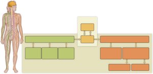
The nervous system is divided into two main parts: the Central Nervous System (CNS) and the Peripheral Nervous System (PNS).
Central Nervous System (CNS): Consists of the brain and spinal cord. It receives, processes, and transfers information.
Peripheral Nervous System (PNS): Composed of nerves outside the CNS. It has sensory and motor divisions, further divided into somatic and autonomic branches.
Neurons: Communication Cells of the Nervous System
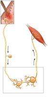
Neurons are specialized cells that generate and conduct electrical impulses, enabling communication within the nervous system. There are three main types of neurons:
Sensory neurons: Receive stimuli and transmit information to the CNS.
Interneurons: Transmit information within the CNS.
Motor neurons: Transmit information from the CNS to effectors (muscles or glands).
Each neuron consists of three parts:
Cell body: Contains the nucleus and most organelles.
Dendrites: Receive incoming information.
Axon: Conducts impulses away from the cell body.
Action Potentials and Resting Potential
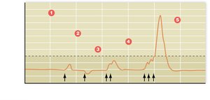
Neurons communicate via action potentials, which are electrical impulses generated when graded potentials reach a threshold. The sodium-potassium pump maintains the resting potential of neurons.
Resting potential: The voltage difference across the cell membrane in a resting neuron, typically –70 mV.
Sodium-potassium pump: Actively transports 3 Na+ out and 2 K+ into the cell, maintaining cell volume and resting potential.
Graded Potentials
Graded potentials are transient changes in the resting potential that may depolarize or hyperpolarize the membrane. Summation of graded potentials can trigger an action potential.
Action Potential Phases
Depolarization: Na+ channels open, Na+ enters, membrane becomes positive.
Repolarization: Na+ channels close, K+ channels open, K+ exits, membrane returns to negative.
Reestablishment: K+ channels close, sodium-potassium pump restores resting potential.
Action potentials are all-or-none and self-propagating, moving like a wave down the axon. The frequency of action potentials encodes stimulus strength.
Neuroglial Cells: Support and Protection
Neuroglial cells, including Schwann cells and oligodendrocytes, support and protect neurons. They form myelin sheaths, which insulate axons and speed up impulse transmission via saltatory conduction.
Schwann cells: Form myelin in the PNS.
Oligodendrocytes: Form myelin in the CNS.
Myelin sheath: Saves energy, speeds transmission, aids regeneration.
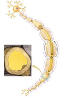
Disorders Associated with Myelin Degeneration
Multiple sclerosis (MS): Progressive damage to CNS myelin.
Amyotrophic lateral sclerosis (ALS): Progressive damage to motor area myelin.
Synaptic Transmission
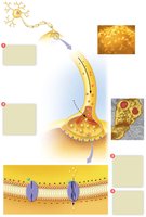
Information is transferred from a neuron to its target (another neuron, muscle, or gland) via synapses. Synaptic transmission involves the release and diffusion of neurotransmitters across the synaptic cleft.
Action potential arrives at axon terminus, Ca2+ enters axon bulb.
Ca2+ triggers neurotransmitter release from vesicles.
Neurotransmitter binds to receptors on postsynaptic membrane, opening gated channels.
Graded potential results from Na+ diffusion.
Neurotransmitters: Excitatory and Inhibitory Effects
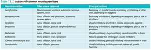
Neurotransmitters can be excitatory (depolarize postsynaptic cell) or inhibitory (hyperpolarize postsynaptic cell). The effect depends on neurotransmitter type, receptor, and ion channel.
Neurotransmitter | Site where released | Principal actions |
|---|---|---|
Acetylcholine | Neuromuscular junctions, autonomic nervous system | Excitatory on skeletal muscles; excitatory or inhibitory at other sites |
Norepinephrine | Areas of brain, spinal cord, autonomic nervous system | Excitatory or inhibitory; plays role in emotions |
Serotonin | Areas of brain, spinal cord | Usually inhibitory; involved in mood, sleep cycle |
Dopamine | Areas of brain, parts of peripheral nervous system | Excitatory or inhibitory; depending on receptor, plays a role in emotions |
Glutamate | Areas of brain, spinal cord | Usually excitatory; major excitatory neurotransmitter in brain |
Endorphins | Areas of brain, spinal cord | Usually inhibitory; natural painkiller; induces euphoria |
Somatostatin | Areas of brain, pancreas | Usually inhibitory; inhibits pancreatic release of growth hormone |
Integration and Processing in Postsynaptic Neurons
Postsynaptic neurons integrate information from multiple synapses. The response depends on the number and type (excitatory or inhibitory) of synapses.
Convergence: One neuron receives input from many others.
Divergence: One neuron sends action potentials to multiple others.
Peripheral Nervous System: Relaying Information
The PNS relays information between tissues and the CNS. Nerves contain axons of many neurons and are wrapped in protective sheaths. Cranial nerves connect to the brain, while spinal nerves connect to the spinal cord.
Somatic Division: Control of Skeletal Muscles
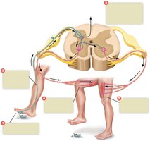
The somatic division controls voluntary movements and reflexes. Reflexes are involuntary responses mediated by the spinal cord and nerves.
Flexor (withdrawal) reflex: Withdraws limb from painful stimulus.
Crossed extensor reflex: Extends opposite limb for balance.
Stretch reflex: Maintains posture and movement.
Autonomic Division: Automatic Body Functions
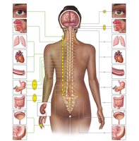
The autonomic division controls involuntary functions of internal organs. It consists of the sympathetic and parasympathetic divisions, which work antagonistically to maintain homeostasis.
Sympathetic division: Prepares body for emergencies (fight-or-flight).
Parasympathetic division: Relaxes body, promotes digestion and rest.
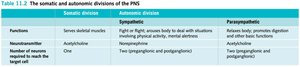
Division | Function | Neurotransmitter | Number of neurons to target |
|---|---|---|---|
Somatic | Serves skeletal muscles | Acetylcholine | One |
Sympathetic | Fight-or-flight | Norepinephrine | Two (preganglionic and postganglionic) |
Parasympathetic | Rest and digest | Acetylcholine | Two (preganglionic and postganglionic) |
CNS: Brain and Spinal Cord
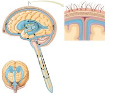
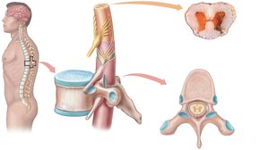
The CNS is protected by bone, meninges, cerebrospinal fluid, and the blood-brain barrier. The spinal cord relays information between the brain and body, with white matter (myelinated tracts) and gray matter (cell bodies and dendrites).
Brain: Processing and Acting on Information
The brain is divided into hindbrain, midbrain, and forebrain, each with distinct functions.
Hindbrain: Medulla oblongata (vital functions), cerebellum (movement coordination), pons (information flow).
Midbrain: Vision, hearing, sleep/wakefulness cycles.
Forebrain: Emotions, conscious thought, homeostasis.
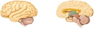
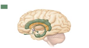
Limbic System and Emotions
The limbic system controls emotional behavior and motivational drives. The hypothalamus serves as a gateway to and from the limbic system.
Cerebrum and Cerebral Cortex
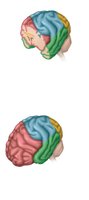
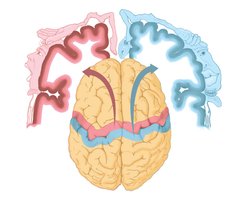
The cerebrum is responsible for higher brain functions. The cerebral cortex is divided into four lobes, each with specific functions:
Occipital lobe: Visual processing.
Temporal lobe: Auditory processing, language comprehension.
Parietal lobe: Sensory information from skin.
Frontal lobe: Motor activity, speech, conscious thought.
Memory
Memory involves storing and retrieving information. Short-term memory is stored in the limbic system, while long-term memory involves permanent changes in the cerebral cortex.
Psychoactive Drugs and Nervous System Disorders
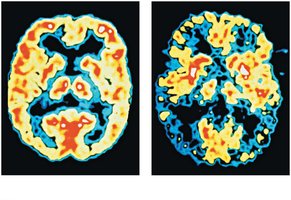
Psychoactive drugs affect consciousness, emotions, and behavior by crossing the blood-brain barrier and altering neurotransmitter activity. Disorders of the nervous system include trauma, infections, and diseases affecting neural and synaptic transmission.
Epilepsy: Abnormal electrical activity (seizures).
Alzheimer’s disease: Progressive memory loss, dementia.
Parkinson’s disease: Degenerative disorder affecting motor activity.