 Back
The Nervous System: Structure, Function, and Disorders
Study Guide - Smart Notes
 Tailored notes based on your materials, expanded with key definitions, examples, and context.
The Nervous System
Overview of the Nervous System
The nervous system is responsible for receiving, processing, storing, and transferring information throughout the body. It is divided into the central nervous system (CNS) and the peripheral nervous system (PNS).
CNS: Composed of the brain and spinal cord; processes and integrates information.
PNS: Consists of nerves and ganglia outside the CNS; subdivided into sensory (afferent) and motor (efferent) divisions.
Somatic Division: Controls voluntary movements of skeletal muscles.
Autonomic Division: Regulates involuntary functions of smooth muscle, cardiac muscle, and glands; includes sympathetic and parasympathetic subdivisions.
Neurons: Structure and Function
Neurons are specialized cells that generate and transmit electrical impulses called action potentials.
Sensory Neurons: Respond to stimuli and transmit information to the CNS.
Interneurons: Located in the CNS; transmit impulses between neurons.
Motor Neurons: Carry impulses from the CNS to effectors (muscles and glands).
Neuron Structure: Includes a cell body (with nucleus and organelles), dendrites (receive signals), and an axon (conducts impulses).
Axon Terminals: Branches at the end of the axon, each with an axon bulb for neurotransmitter release.
Action Potential: Generation and Propagation
Action potentials are the primary means of long-distance communication in the nervous system.
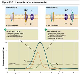
Resting Potential: The neuron maintains a voltage of -70 mV across its membrane, due to the sodium-potassium pump and negative molecules in the interstitial fluid.
Graded Potentials: Local changes in membrane potential; can be depolarizing (closer to zero) or hyperpolarizing (more negative).
Threshold: If graded potentials reach a certain level, an action potential is triggered.
Depolarization: Sodium channels open, Na+ enters the cell, membrane potential becomes positive.
Repolarization: Sodium channels close, potassium channels open, K+ exits the cell, membrane potential returns to negative.
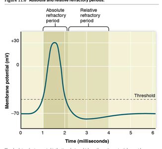
Refractory Periods:
Absolute Refractory Period: No new action potential can be generated.
Relative Refractory Period: A stronger stimulus is required to generate an action potential.
All-or-None Principle: Action potentials either occur fully or not at all, and always propagate in one direction.
Neuroglial Cells: Support and Protection
Neuroglial cells provide structural and functional support to neurons.
Schwann Cells: Produce myelin sheath in the PNS, which insulates axons and speeds up impulse transmission.
Nodes of Ranvier: Gaps between myelin sheaths where action potentials jump, increasing speed (saltatory conduction).
Astrocytes: Maintain chemical environment in the CNS.
Oligodendrocytes: Form myelin sheaths in the CNS.
Diseases:
Multiple Sclerosis: Damage to oligodendrocyte sheaths disrupts CNS transmission.
Amyotrophic Lateral Sclerosis (ALS): Progressive weakening of skeletal muscles due to spinal cord damage.
Synaptic Transmission and Neurotransmitters
Information is transferred from neuron to target via synaptic transmission, involving neurotransmitter release.
Presynaptic Membrane: Releases neurotransmitters into the synaptic cleft.
Postsynaptic Membrane: Receives neurotransmitters, leading to graded potentials.
Excitatory Neurotransmitters: Depolarize postsynaptic cell (e.g., glutamate).
Inhibitory Neurotransmitters: Hyperpolarize postsynaptic cell (e.g., GABA).
Examples:
Acetylcholine: Excites skeletal muscles.
Norepinephrine: Excites or inhibits, involved in emotions.
Serotonin: Inhibitory, affects mood, sleep, appetite.
Dopamine: Excites or inhibits, involved in emotion.
Endorphins: Inhibitory, natural pain relief.
Neurotransmitter Fate: Reuptake, enzymatic destruction, or diffusion away.
Integration and Processing of Information
Postsynaptic neurons integrate multiple inputs, determining whether threshold is reached for action potential generation.
Convergence: One neuron receives input from many others.
Divergence: One neuron sends output to many others.
Nerves and Signal Transmission
Nerves are bundles of axons transmitting signals to and from the CNS.
Cranial Nerves: 12 pairs connect to the brain.
Spinal Nerves: 31 pairs connect to the spinal cord via dorsal (sensory) and ventral (motor) roots.
Somatic Division: Controls voluntary skeletal muscle movements and reflexes.
Autonomic Division: Controls involuntary functions; requires two neurons (preganglionic and postganglionic).
Sympathetic Division: Prepares body for 'fight or flight'; uses norepinephrine.
Parasympathetic Division: Promotes 'rest and digest'; uses acetylcholine.
Central Nervous System: Brain and Spinal Cord
The CNS is protected by bone, meninges, cerebrospinal fluid, and the blood-brain barrier.
Meninges: Three layers (dura mater, arachnoid, pia mater) protect the CNS.
Cerebrospinal Fluid: Cushions and absorbs shock.
Blood-Brain Barrier: Regulates passage of substances into brain tissue.
Spinal Cord: Relays information between brain and body; contains white matter (myelinated axons) and gray matter (cell bodies and unmyelinated axons).
Brain Structure and Function
The brain is divided into hindbrain, midbrain, and forebrain, each with specialized functions.
Hindbrain: Coordinates basic, automatic, and vital tasks (medulla oblongata, cerebellum, pons).
Midbrain: Coordinates muscle groups and responses to visual and auditory stimuli.
Forebrain: Integrates sensory input and determines complex behaviors (hypothalamus, thalamus, limbic system, cerebrum).
Limbic System: Involved in emotion and memory; hippocampus stores memories.
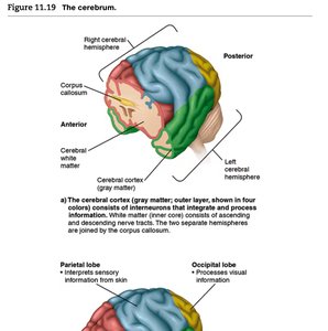
Cerebrum: Responsible for language, decision making, conscious thought; divided into left and right hemispheres connected by the corpus callosum.
Cerebral Cortex: Outer layer of gray matter; integrates sensory information, stores memory, and controls skeletal muscles.
Lobes:
Occipital Lobe: Visual information.
Temporal Lobe: Auditory information, language, perceptual judgment.
Frontal Lobe: Motor activity, speech, conscious thought.
Parietal Lobe: Somatosensory information.
Memory: Storage and Retrieval
Memory involves both short-term and long-term processes.
Short-Term Memory: Stored in the limbic system; lasts for hours.
Long-Term Memory: Transferred to the hippocampus and cerebral cortex; lasts for days or years.
Neuronal Changes: Long-term memory involves structural changes in neurons.
Psychoactive Drugs and Brain Function
Psychoactive drugs alter consciousness, emotions, and behavior by affecting neurotransmitter concentrations or actions.
Cocaine: Blocks dopamine reuptake, leading to addiction.
Opioids: Bind to endorphin receptors; naloxone can block their effects.
Amphetamines: Used to treat ADHD.
Tolerance: Leads to addiction.
Disorders of the Nervous System
The nervous system can be affected by trauma, infection, and degenerative diseases.
Trauma: Includes concussion, spinal cord injury (paraplegia, quadriplegia).
Encephalitis: Inflammation of the brain, often viral.
Meningitis: Inflammation of the meninges.
Rabies: Viral infection affecting the brain.
Brain Tumors: Increase intracranial pressure, disrupt function.
Multiple Sclerosis and ALS: Disorders of neural transmission.
Epilepsy: Abnormal brain cell activity, seizures.
Alzheimer's Disease: Dementia, abnormal protein deposits, loss of acetylcholine neurotransmission.
Parkinson's Disease: Degeneration of dopamine-releasing neurons, affects motor activity.
Electroencephalogram (EEG) and Sleep
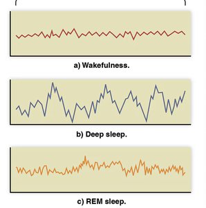
EEG measures electrical activity in the brain, which varies with wakefulness and sleep stages.
Wakefulness: Low amplitude, high frequency waves.
Deep Sleep: High amplitude, low frequency waves.
REM Sleep: Mixed frequency, associated with dreaming.
Key Equations and Concepts
Resting Membrane Potential:
Action Potential Threshold:
Sodium-Potassium Pump: per ATP hydrolyzed
Summary Table: Major Neurotransmitters
Neurotransmitter | Location | Principal Action |
|---|---|---|
Acetylcholine | Neuromuscular junctions, CNS | Excites skeletal muscles |
Norepinephrine | Brain, spinal cord, autonomic NS | Excites/inhibits, emotions |
Serotonin | Brain, spinal cord | Inhibitory, mood, sleep, appetite |
Dopamine | Brain, PNS | Excites/inhibits, emotion |
Glutamate | Brain, spinal cord | Excitatory, major in brain |
Endorphins | Brain, spinal cord | Inhibitory, pain relief |
GABA | Brain, spinal cord | Inhibitory, principal in brain |
Somatostatin | Brain, pancreas | Inhibitory, inhibits growth hormone |
Additional info: Academic context and expanded explanations were added to clarify mechanisms, terminology, and clinical relevance for Human Biology students.
