 Back
A Tour of the Cell: Structure and Function in Eukaryotic and Prokaryotic Cells
Study Guide - Smart Notes
 Tailored notes based on your materials, expanded with key definitions, examples, and context.
A Tour of the Cell
Introduction to Cell Structure and Function
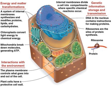
Cells are the fundamental units of life, and their internal organization enables them to carry out essential biological functions. Eukaryotic cells, in particular, possess complex internal membranes and organelles that compartmentalize cellular processes, allowing for greater efficiency and specialization.
Studying Cells: Microscopy and Biochemistry
Microscopy Techniques
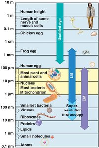
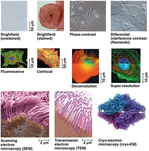
Biologists use various types of microscopes to visualize cells and their components, as most cells are too small to be seen with the naked eye. Light microscopes (LM) use visible light and glass lenses to magnify specimens up to 1,000 times, while electron microscopes (EM) provide much higher resolution, allowing for the study of organelles.
Light Microscopy: Includes brightfield, phase-contrast, fluorescence, and confocal techniques.
Electron Microscopy: Includes scanning (SEM), transmission (TEM), and cryo-electron microscopy.
Resolution: The ability to distinguish two close objects as separate; EM has much higher resolution than LM.
Cell Fractionation
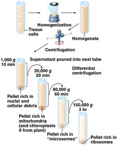
Cell fractionation is a technique used to separate cellular components based on size and density, typically using centrifugation. This allows scientists to study the functions of individual organelles.
Homogenization: Breaking open cells to release their contents.
Differential Centrifugation: Sequentially spinning homogenate at increasing speeds to separate nuclei, mitochondria, microsomes, and ribosomes.
Prokaryotic vs. Eukaryotic Cells
Basic Features of All Cells
Both prokaryotic and eukaryotic cells share certain features:
Plasma membrane: Selective barrier for transport.
Cytosol: Semifluid substance inside the cell.
Chromosomes: Carry genetic information.
Ribosomes: Sites of protein synthesis.
Prokaryotic Cells
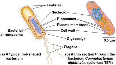
Prokaryotic cells (Bacteria and Archaea) lack a nucleus and membrane-bound organelles. Their DNA is located in the nucleoid region, and the cytoplasm is bound by the plasma membrane.
No nucleus
No membrane-bound organelles
Cell wall, fimbriae, flagella, and glycocalyx may be present
Eukaryotic Cells
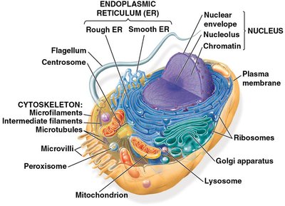
Eukaryotic cells (protists, fungi, animals, plants) have a nucleus enclosed by a double membrane and numerous membrane-bound organelles. They are generally larger than prokaryotic cells.
Nucleus: Contains DNA
Membrane-bound organelles: Specialized for different functions
Cytoplasm: Region between plasma membrane and nucleus
Cell Size and Surface Area-to-Volume Ratio
Importance of Surface Area-to-Volume Ratio
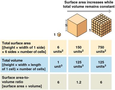
The efficiency of a cell depends on its surface area-to-volume ratio. As cells grow, their volume increases faster than their surface area, limiting the rate of transport across the membrane.
High surface area-to-volume ratio: More efficient transport and metabolic activity
Formula:
The Plasma Membrane
Structure and Function
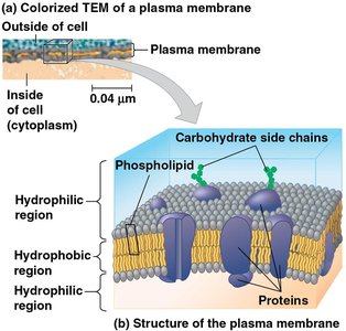
The plasma membrane is a selective barrier composed of a phospholipid bilayer with embedded proteins and carbohydrate side chains. It regulates the passage of oxygen, nutrients, and waste.
Phospholipid bilayer: Hydrophilic heads face outward, hydrophobic tails inward
Proteins: Facilitate transport and communication
Carbohydrate chains: Cell recognition
Internal Organization of Eukaryotic Cells
Compartmentalization and Organelles
Eukaryotic cells are divided into compartments by internal membranes, allowing incompatible processes to occur simultaneously. The basic fabric of biological membranes is a double layer of phospholipids.
Organelles: Nucleus, endoplasmic reticulum, Golgi apparatus, lysosomes, vacuoles, mitochondria, chloroplasts
Plant vs. Animal Cells: Most organelles are shared, but plant cells have chloroplasts and a central vacuole
The Nucleus and Ribosomes
Nucleus: Information Central
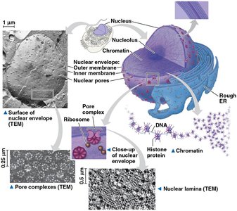
The nucleus houses most of the cell's genetic material and is surrounded by a double membrane called the nuclear envelope. Nuclear pores regulate the entry and exit of molecules.
Chromosomes: DNA molecules associated with proteins (chromatin)
Nucleolus: Site of ribosomal RNA synthesis
Ribosomes: Protein Factories
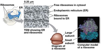
Ribosomes are complexes of ribosomal RNA and protein, responsible for protein synthesis. They can be free in the cytosol or bound to the endoplasmic reticulum or nuclear envelope.
Free ribosomes: Synthesize proteins for use within the cell
Bound ribosomes: Synthesize proteins for secretion or membrane insertion
The Endomembrane System
Components and Functions
The endomembrane system regulates protein traffic and performs metabolic functions. It includes the nuclear envelope, endoplasmic reticulum (ER), Golgi apparatus, lysosomes, vacuoles, and plasma membrane.
Continuous or connected via vesicles
Endoplasmic Reticulum (ER)
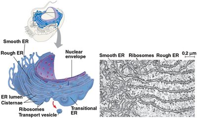
The ER is a biosynthetic factory, consisting of smooth and rough regions. The smooth ER synthesizes lipids, detoxifies drugs, and stores calcium ions. The rough ER, studded with ribosomes, synthesizes proteins and distributes transport vesicles.
Smooth ER: Lipid synthesis, detoxification, calcium storage
Rough ER: Protein synthesis, glycoprotein production, membrane factory
Golgi Apparatus
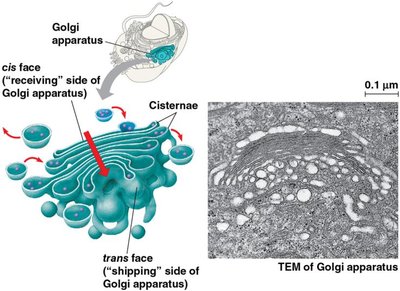
The Golgi apparatus is the shipping and receiving center of the cell, consisting of flattened sacs called cisternae. It modifies, sorts, and packages products from the ER into transport vesicles.
Cis face: Receiving side
Trans face: Shipping side
Lysosomes
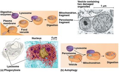
Lysosomes are digestive compartments containing hydrolytic enzymes. They digest macromolecules, recycle cellular components (autophagy), and fuse with food vacuoles formed by phagocytosis.
Acidic environment: Optimal for enzyme activity
Autophagy: Recycling of organelles
Vacuoles
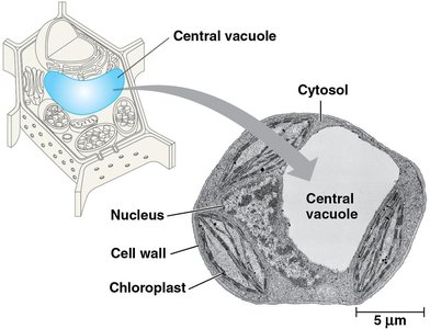
Vacuoles are large vesicles with diverse functions. Food vacuoles form by phagocytosis, contractile vacuoles expel excess water, and central vacuoles in plants store ions and contribute to cell growth.
Central vacuole: Main repository of inorganic ions in plant cells
Mitochondria and Chloroplasts: Energy Conversion
Endosymbiont Theory
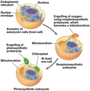
Mitochondria and chloroplasts are organelles that convert energy from one form to another. The endosymbiont theory proposes that these organelles originated from prokaryotic cells engulfed by ancestral eukaryotes.
Double membrane
Own DNA and ribosomes
Reproduce independently
Mitochondria
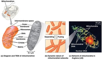
Mitochondria are the sites of cellular respiration, generating ATP. They have a smooth outer membrane and a highly folded inner membrane (cristae), creating compartments for metabolic reactions.
Intermembrane space
Mitochondrial matrix
Cristae: Increase surface area for ATP synthesis
Summary Table: Comparison of Prokaryotic and Eukaryotic Cells
Feature | Prokaryotic Cells | Eukaryotic Cells |
|---|---|---|
Nucleus | No | Yes |
Membrane-bound organelles | No | Yes |
Size | Small (0.1–5 µm) | Larger (10–100 µm) |
DNA location | Nucleoid | Nucleus |
Cell wall | Usually present | Plants and fungi only |
Examples | Bacteria, Archaea | Protists, fungi, animals, plants |
Conclusion
The internal organization of cells, especially eukaryotic cells, is essential for the performance of life functions. Understanding cell structure, organelles, and their functions provides a foundation for studying more advanced topics in biology.
