 Back
ch 40 How Animals Sense Their Environment: The Senses
Study Guide - Smart Notes
 Tailored notes based on your materials, expanded with key definitions, examples, and context.
The Senses
Overview of Sensory Systems
Animals rely on specialized sensory systems to detect and interpret both external and internal stimuli. Sensory receptors are specialized cells or nerve endings that convert environmental signals into electrical impulses for the nervous system. These receptors are classified based on the type of stimulus they detect.
Sensory Receptors: Specialized cells that detect environmental stimuli and convert them into electrical signals.
Categories: Mechanoreceptors, thermoreceptors, photoreceptors, chemoreceptors, and nociceptors.
Sense Organs: Complex structures (e.g., eyes, ears) that combine receptors with supporting tissues to enhance detection.
Electrical Signaling in Sensory Receptors
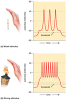
Sensory receptors generate electrical signals called receptor potentials when stimulated. If the signal crosses a threshold, it triggers action potentials that are sent to the brain.
Action Potential Frequency: The intensity of a stimulus is encoded by the frequency of action potentials.
Example: A weak stimulus produces fewer action potentials, while a strong stimulus produces more frequent action potentials.
Sensing Temperature
Thermoreceptors
Thermoreceptors are specialized sensory cells that detect changes in temperature.
Skin Thermoreceptors: Respond to heat or cold and help maintain body temperature.
Brain Thermoreceptors: Monitor core temperature and trigger physiological responses.
Firing Patterns: Cold receptors fire more rapidly below 77°F; warm receptors fire more rapidly above 91°F.
Detecting Mechanical Stimuli
Mechanoreceptors
Mechanoreceptors respond to physical deformation such as touch, pressure, vibration, and stretch.
Skin Mechanoreceptors: Detect touch, vibration, and pressure.
Stretch Receptors: Found in internal organs (e.g., intestines, stomach, bladder) and muscles.
Inner Ear Mechanoreceptors: Respond to sound, gravity, and movement.
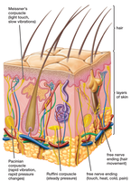
Density Variation: Fingertips have many mechanoreceptors; the back has few.
Animal Examples: Spiders detect web vibrations; fish use lateral lines to sense water movement.
Hearing: How Animals Detect Sound
Anatomy of the Ear
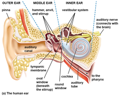
The mammalian ear is divided into three main parts: outer, middle, and inner ear.
Outer Ear: Pinna collects sound; auditory canal directs it to the eardrum.
Middle Ear: Tympanic membrane and three bones (malleus, incus, stapes) transmit vibrations; auditory tube equalizes pressure.
Inner Ear: Cochlea converts vibrations into electrical signals; vestibular system detects balance and movement.
Sound Detection and Signal Transmission
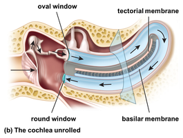
Cochlea: Fluid movement vibrates the basilar membrane, stimulating hair cells.
Hair Cells: Release neurotransmitters onto neurons, forming the auditory nerve.
Action Potentials: Signals travel to the brain's auditory centers.
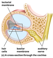
Sound Properties: Loudness and Pitch
Loudness: Greater hair-cell bending and more frequent signals indicate louder sounds; very loud sounds can damage hair cells.
Pitch: Different sound frequencies vibrate different parts of the basilar membrane; high pitches near the oval window, low pitches near the cochlea’s tip.
Sensing Gravity and Movement
Vestibular Apparatus
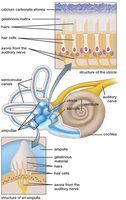
The vestibular apparatus in the inner ear detects gravity, head position, and movement.
Utricle and Saccule: Gravity shifts calcium carbonate stones, bending hair cells to signal head tilt.
Semicircular Canals: Three fluid-filled canals detect movement in different directions; head acceleration moves fluid, bending hair cells.
Neurotransmitter Release: Bent hair cells release neurotransmitters, triggering nerve signals to the brain’s balance centers.
Vision: How Animals Detect Light
Types of Eyes and Photoreceptors
Most animals detect light, ranging from simple light/dark detection to complex image formation.
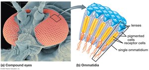
Compound Eyes: Found in arthropods; composed of ommatidia, excellent at detecting movement.
Image-Forming Eyes: Found in cephalopods and vertebrates; use photoreceptors to convert light into electrical signals.
Human Eye Structure
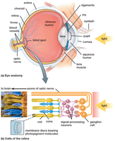
The human eye contains over 100 million photoreceptors and functions similarly to a camera.
Accessory Structures: Maintain eye shape, control light, and focus it.
Retina: Photoreceptors convert light into electrical signals.
How Light Enters and Is Focused
Sclera: Protects the eyeball.
Cornea: Entry point for light; passes through aqueous humor and pupil.
Iris: Regulates pupil size and incoming light.
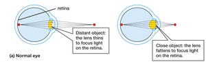
Lens: Focuses light onto the retina.
Vitreous Humor: Maintains eye shape.
Focusing and Seeing
Fovea: Area of sharpest vision; muscles adjust lens shape for focusing.
Choroid: Nourishes retina and absorbs stray light.
Photoreceptors: Rods and Cones
Rods: Highly sensitive to light; support dim-light vision but not color.
Cones: Three types sensitive to red, green, or blue; enable color vision.
Distribution: Day-active vertebrates have more cones; nocturnal species have more rods.
Vision Adaptations
Binocular Vision: Forward-facing eyes provide depth perception for predators and omnivores.
Wide Field of View: Side-facing eyes in herbivores help detect predators.
Chemical Senses: Smell and Taste
Chemoreceptors
Chemoreceptors detect chemicals inside or outside the body, including blood oxygen and glucose levels.
Olfaction (Smell): Humans have 5–10 million olfactory neurons with about 400 receptor types; each neuron is tuned to specific odor molecules.
Gustation (Taste): The tongue has about 5,000 taste buds, many embedded in papillae.
Taste Buds and Taste Types
Taste Buds: Contain receptor, supporting, and stem cells; microvilli detect dissolved chemicals.
Stem Cells: Replace worn or damaged taste cells.
Five Tastes: Sour, salty, sweet, bitter, and umami (savory).
Mechanisms: Sour and salty involve ions entering cells; sweet, bitter, and umami involve molecules binding to receptors.
Flavor Perception
Flavor: Combines taste and smell; foods activate multiple taste receptors and release aromas detected by olfactory receptors.
Example: Food tastes bland when smell is blocked (e.g., holding your nose).
Sensing Pain
Pain Receptors (Nociceptors)
Pain receptors detect harmful stimuli such as extreme temperatures, excessive stretching, or injury-related chemicals.
Function: Send signals to the brain interpreted as pain, helping protect the body from further harm.
Principal Categories of Vertebrate Sensory Receptors
Table: Sensory Receptor Types
Receptor Type | Stimulus Detected | Example Location |
|---|---|---|
Mechanoreceptor | Touch, pressure, vibration, stretch | Skin, muscles, inner ear |
Thermoreceptor | Temperature changes | Skin, brain |
Photoreceptor | Light | Eyes (retina) |
Chemoreceptor | Chemicals (taste, smell, blood chemistry) | Tongue, nose, blood vessels |
Nociceptor | Painful stimuli | Skin, internal organs |
Additional info: Table entries inferred from textbook context and standard biology knowledge.
