 Back
The Central Nervous System: Structure, Development, and Function
Study Guide - Smart Notes
 Tailored notes based on your materials, expanded with key definitions, examples, and context.
The Central Nervous System
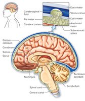
Structural Organization of the Brain
The central nervous system (CNS) consists of the brain and spinal cord, serving as the main processing center for sensory input and motor output. It is responsible for integrating information, maintaining homeostasis, and supporting higher functions such as learning, memory, and consciousness.
Brain: The primary organ for processing and integrating information.
Spinal Cord: Connects the brain to the peripheral nervous system and mediates reflexes.
Interneurons: Integrate sensory information and direct appropriate responses.
Consciousness: Encompasses perception, learning, memory, emotions, and self-awareness.
Embryonic Development of the CNS
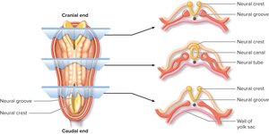
The CNS originates from the ectoderm during embryonic development. Around 20 days after conception, a neural groove forms, which becomes the neural tube—the precursor to the CNS. The neural crest, located between the neural tube and developing epidermis, forms the peripheral nervous system (PNS) ganglia.
Neural Tube: Develops into the brain and spinal cord.
Neural Crest: Forms PNS ganglia.
Developmental Sequence of the Brain
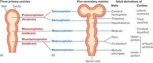
During the fourth week of embryonic development, three primary brain vesicles form:
Prosencephalon: Forebrain
Mesencephalon: Midbrain
Rhombencephalon: Hindbrain
By the fifth week, these vesicles differentiate into five regions:
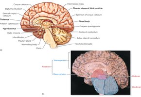
Telencephalon: Cerebral hemispheres, lateral ventricles
Diencephalon: Thalamus, hypothalamus, third ventricle
Mesencephalon: Midbrain, cerebral aqueduct
Metencephalon: Pons, cerebellum, upper fourth ventricle
Myelencephalon: Medulla oblongata, lower fourth ventricle
Ventricles and Cerebrospinal Fluid
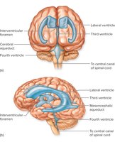
The brain contains four ventricles filled with cerebrospinal fluid (CSF), which cushions the brain, removes waste, and maintains chemical stability. The choroid plexus produces CSF, which circulates through the ventricles and central canal of the spinal cord, eventually being reabsorbed into the blood.
CSF Pathway: Lateral ventricles → third ventricle → cerebral aqueduct → fourth ventricle → central canal/subarachnoid space
Choroid Plexus: Epithelial cells and capillaries that produce CSF
Blood-Brain Barrier: Tight junctions in the choroid plexus prevent unwanted substances from entering the CNS
Brain Statistics and Neurogenesis
Gray Matter: Cell bodies and dendrites; forms cortex and deep nuclei
White Matter: Myelinated axons; forms tracts
Adult Brain: ~100 billion neurons, weighs 1.5 kg, receives 15% of blood flow
Neurogenesis: Formation of new neurons from neural stem cells occurs in adults, especially in the hippocampus and lateral ventricles
The Cerebrum
Introduction and Structure
The cerebrum, derived from the telencephalon, is the largest portion of the brain and is responsible for higher mental functions. It consists of two hemispheres connected by the corpus callosum.
Cerebral Cortex: Outer region of gray matter (2–4 mm thick)
Gyri: Raised folds
Sulci: Depressed grooves
Convolutions: Gyri and sulci together
Lobes: Frontal, Parietal, Temporal, Occipital, Insula
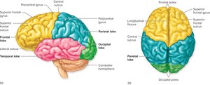
Functional Regions of the Cerebral Cortex
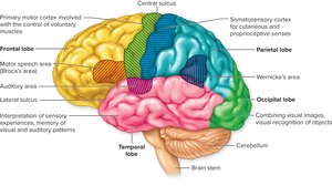
Each lobe of the cerebrum has specialized functions.
Frontal Lobe: Motor control, speech production (Broca's area)
Parietal Lobe: Somatosensory processing
Temporal Lobe: Auditory centers, memory, interpretation
Occipital Lobe: Visual processing
Insula: Memory encoding, integration of sensory and visceral responses
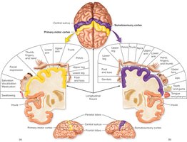
Frontal and Parietal Lobes: Motor and Sensory Maps
The precentral gyrus (frontal lobe) is the primary motor cortex, while the postcentral gyrus (parietal lobe) is the primary somatosensory cortex. Both regions are mapped according to the density of receptors and motor innervations, not body part size.
Motor Cortex: Controls voluntary movements
Somatosensory Cortex: Processes sensory input from skin, muscles, joints
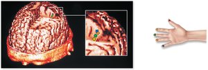
Brain Imaging and Visualization
Modern imaging techniques allow visualization of brain structure and function.
CT: X-ray absorption for soft tissue
PET: Radioisotope tracers for metabolic activity
MRI: Magnetic alignment of protons for tissue contrast
fMRI: Blood flow changes for neuronal activity
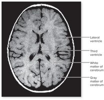
Electroencephalogram (EEG) and Brain Waves
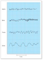
EEG measures electrical activity in the cerebral cortex. Four main wave patterns are observed:
Alpha Waves: Awake, relaxed
Beta Waves: Mental activity, visual stimulation
Theta Waves: Sleep, attention, memory tasks
Delta Waves: Deep sleep, brain damage if seen in awake adults
Basal Nuclei and Motor Circuits
Basal Nuclei Structure and Function
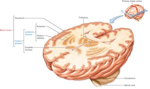
Basal nuclei (basal ganglia) are masses of gray matter deep in the cerebrum, involved in voluntary movement control.
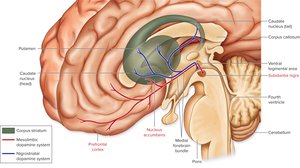
Corpus Striatum: Caudate nucleus, putamen, globus pallidus
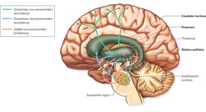
Motor Circuit: Involves excitatory and inhibitory neurotransmitters (glutamate, GABA, dopamine)
Cerebral Lateralization and Language
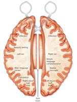
Cerebral Lateralization
Each hemisphere of the brain specializes in certain functions.
Right Hemisphere: Visuospatial tasks, face recognition, music, spatial concepts
Left Hemisphere: Language, analytical ability, speech, writing, calculations
Language Areas
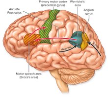
Broca's Area: Motor speech production
Wernicke's Area: Language comprehension
Angular Gyrus: Integration of auditory, visual, and somatosensory information
Limbic System and Emotion
Limbic System Structure
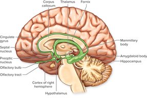
The limbic system is a group of interconnected brain regions responsible for emotional states and memory.
Components: Cingulate gyrus, amygdaloid body, hippocampus, septal nuclei, anterior insula, hypothalamus, thalamus
Papez Circuit: Pathway connecting hippocampus, hypothalamus, thalamus, cingulate gyrus
Memory and Synaptic Plasticity
Types of Memory
Memory is classified into short-term, working, and long-term types. Long-term memory requires structural changes in neurons, including gene activation and protein synthesis.
Nondeclarative (Implicit): Skills and conditioning
Declarative (Explicit): Facts and events (semantic and episodic)
Long-Term Potentiation (LTP)
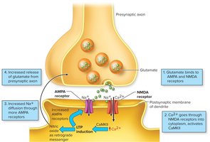
LTP is a process that strengthens synaptic connections, crucial for memory formation.
Glutamate: Binds to AMPA and NMDA receptors
Ca2+ Entry: Activates CaMKII, increases AMPA receptors
Synaptic Strength: Enhanced by increased glutamate release and receptor sensitivity
Pyramidal Cells and Synaptic Changes
Pyramidal cells in the hippocampus grow dendritic spines and increase AMPA receptors during LTP, further strengthening synaptic connections. 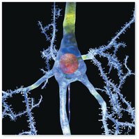
Diencephalon, Midbrain, and Hindbrain
Diencephalon
The diencephalon includes the thalamus, hypothalamus, epithalamus, and part of the pituitary gland.
Thalamus: Relay center for sensory information
Epithalamus: Contains choroid plexus and pineal gland (melatonin secretion)
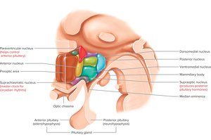
Hypothalamus: Regulates homeostasis, autonomic functions, hormone secretion, circadian rhythms
Midbrain and Hindbrain
Midbrain: Visual and auditory reflexes, motor coordination, reward pathways
Hindbrain: Pons and cerebellum (motor learning, balance), medulla oblongata (autonomic regulation)
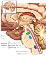
Reticular Activating System (RAS)
The RAS regulates sleep and arousal by tuning sensory stimuli and modulating alertness. 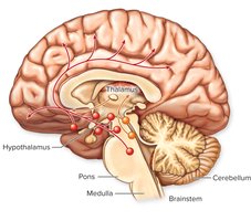
Spinal Cord Tracts and Nerves
Spinal Cord Structure
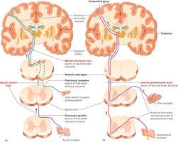
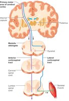
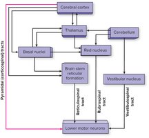
The spinal cord consists of white matter (ascending and descending tracts) surrounding a gray matter core.
Ascending Tracts: Carry sensory information to the brain
Descending Tracts: Carry motor commands from the brain
Cranial and Spinal Nerves
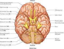
Cranial Nerves: 12 pairs, arise from the brain, mostly mixed sensory and motor
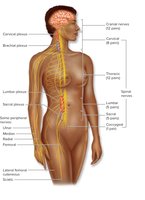
Spinal Nerves: 31 pairs, arise from the spinal cord, all mixed
Reflex Arc
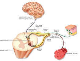
Reflex arcs are unconscious motor responses to sensory stimuli, involving sensory receptors, neurons, interneurons, and effectors.
Somatic Reflex: Skeletal muscle effectors
Autonomic Reflex: Smooth muscle, cardiac muscle, or gland effectors
Additional info:
The notes include expanded academic context and explanations for clarity and completeness, suitable for introductory biology students.
