 Back
39. The Nervous System: Structure, Function, and Organization
Study Guide - Smart Notes
 Tailored notes based on your materials, expanded with key definitions, examples, and context.
The Nervous System
The Nerve Cells
The nervous system is composed of two principal cell types: neurons and glia. Neurons are specialized for receiving, processing, and transmitting information, as well as controlling body movements. Glia assist neuronal function by providing nutrients, regulating the composition of interstitial fluid, protecting against infection, repairing damage, fine-tuning communication, and speeding up electrical signal movement.
Neurons: Receive, process, and transmit information; control body movements.
Glia: Support neurons through various functions including nutrient supply and protection.
Structure of a Typical Neuron
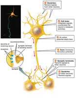
A typical neuron consists of four major structures: dendrites, cell body, axon, and synaptic terminals. Each part has a specialized function in information processing and transmission.
Dendrites: Receive signals from other neurons or sensory stimuli.
Cell Body: Integrates incoming signals and supports metabolic activities.
Axon: Conducts action potentials away from the cell body.
Synaptic Terminals: Transmit signals to other neurons, muscle cells, or gland cells.
Neurotransmitters
Neurotransmitters are chemical messengers released at synapses. They bind to receptors on the receiving cell, causing changes in voltage and influencing whether the neuron will fire an action potential.
Neurotransmitter | Main Function |
|---|---|
Acetylcholine | Stimulates muscle contraction |
Dopamine | Regulates mood, reward, and movement |
Serotonin | Regulates mood, appetite, and sleep |
Norepinephrine | Prepares body for action (fight or flight) |
Glutamate | Main excitatory neurotransmitter |
GABA | Main inhibitory neurotransmitter |
Additional info: | Table entries inferred from standard biology sources |
How Neurons Produce and Transmit Information
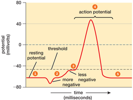
Neurons use electrical signals within the cell and neurotransmitters to pass signals between cells. At rest, neurons maintain a resting potential, with the inside being negative relative to the outside. Stimulation can change this voltage; if it reaches a threshold, an action potential is generated.
Resting Potential: Typically ranges from −40 to −90 mV.
Action Potential: Rapid electrical signal that travels along the axon.
Threshold: The voltage at which an action potential is triggered.
Conduction of Action Potentials
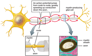
Action potentials travel along the axon without losing strength. Thicker axons conduct faster, and myelin insulation greatly increases speed by allowing the action potential to jump from node to node (saltatory conduction).
Myelin Sheath: Fatty insulation produced by glial cells.
Nodes of Ranvier: Gaps in myelin where action potentials are regenerated.
Synaptic Transmission
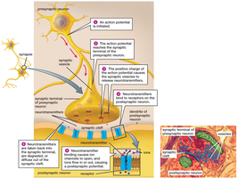
At the synapse, the action potential triggers the release of neurotransmitters from the presynaptic neuron. These chemicals cross the synaptic cleft and bind to receptors on the postsynaptic neuron, causing a postsynaptic potential (PSP).
Excitatory PSP (EPSP): Makes the neuron less negative, increasing the likelihood of firing.
Inhibitory PSP (IPSP): Makes the neuron more negative, decreasing the likelihood of firing.
Integration: The neuron sums EPSPs and IPSPs to determine if threshold is reached.
How the Nervous System Processes Information and Controls Behavior
The nervous system processes information, stores memories, and controls behavior through specialized neurons and interconnected neural networks. Most behaviors are controlled by pathways composed of sensory neurons, interneurons, motor neurons, and effectors.
Sensory Neurons: Detect internal and external stimuli.
Interneurons: Integrate sensory input and memories.
Motor Neurons: Activate muscles or glands.
Effectors: Muscles or glands that perform the response.
Encoding Stimulus Intensity
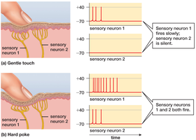
Stimulus intensity is encoded by the frequency of action potentials. Stronger stimuli produce more frequent signals, while individual action potentials remain the same size.
Organization of the Nervous Systems
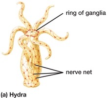
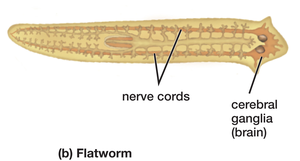
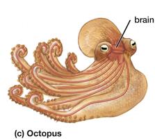
Animals exhibit two main types of nervous system organization: diffuse nerve networks and centralized nervous systems. Cnidarians, such as Hydra, have a nerve net with ganglia, while bilaterally symmetrical animals have centralized nervous systems with brains and nerve cords.
Diffuse Nerve Networks: Found in radially symmetrical animals (e.g., Hydra).
Centralized Nervous Systems: Found in complex, bilaterally symmetrical animals.
The Human Nervous System
The human nervous system is divided into the central nervous system (CNS) and the peripheral nervous system (PNS). The CNS includes the brain and spinal cord, while the PNS consists of nerves and neurons outside the CNS.
CNS: Processes sensory information, supports thought, and directs responses.
PNS: Connects CNS to the body; contains sensory neuron cell bodies and neurons controlling involuntary functions.
Peripheral Nerves
Sensory Neurons: Carry information to the CNS.
Motor Neurons: Innervate skeletal muscles for voluntary movement.
Autonomic Neurons: Control involuntary movements and activities.
Somatic and Autonomic Nervous Systems
Somatic Nervous System: Controls voluntary movement.
Autonomic Nervous System: Controls involuntary functions; divided into sympathetic (fight or flight) and parasympathetic (rest and digest) divisions.
Protection and Function of the CNS
Protection: Skull, vertebrae, meninges, and cerebrospinal fluid (CSF).
Blood–Brain Barrier: Seals brain capillaries, protecting from harmful chemicals.
Spinal Cord: Controls reflexes and carries signals between brain and body.
Spinal Cord Injury
Can block signals between brain and body below the injury, causing loss of sensation and movement.
Additional info: Table of neurotransmitters and some details inferred from standard biology textbooks for completeness.
