 Back
Chapter 17: Radioactivity and Nuclear Chemistry – Study Notes
Study Guide - Smart Notes
 Tailored notes based on your materials, expanded with key definitions, examples, and context.
Radioactivity and Nuclear Chemistry
Introduction to Radioactivity
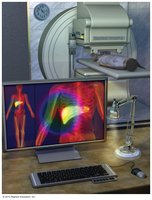
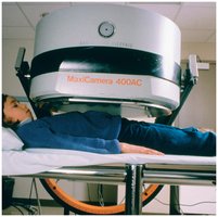
Radioactivity is a fundamental concept in nuclear chemistry, involving the emission of energetic particles from the nuclei of unstable atoms. This process is harnessed in nuclear medicine to obtain clear images of internal organs and diagnose diseases such as appendicitis.
Radioactivity is the spontaneous emission of particles or energy from unstable atomic nuclei.
Used in medical imaging to detect abnormalities in organs.
Radioactively tagged antibodies can accumulate in infected areas, allowing detection via emitted radiation.
Nuclear medicine utilizes radioactivity for diagnostic purposes.
The Discovery of Radioactivity
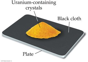
The phenomenon of radioactivity was first discovered by Antoine-Henri Becquerel in 1896. He observed that uranium-containing crystals could expose photographic plates even in the absence of light, leading to the identification of uranic rays.
Becquerel's experiment demonstrated that uranium was the source of emissions, not phosphorescence.
Marie Curie expanded on this work, discovering polonium and radium, both highly radioactive elements.
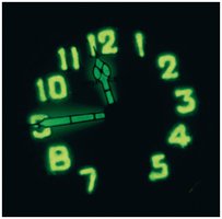
Radium was used in luminous paints for watch dials due to its intense radioactivity.
Curie received Nobel Prizes in both physics and chemistry for her contributions.

Types of Radioactivity
Alpha, Beta, Gamma, and Positron Decay
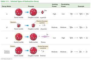
Radioactive emissions are classified into several types, each with distinct properties and effects. The stability of atomic nuclei depends on their size and neutron-to-proton ratio.
Alpha (α) rays: Emission of 2 protons and 2 neutrons.
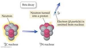
Beta (β) rays: Emission of electrons when a neutron turns into a proton.
Gamma (γ) rays: High-energy photons, electromagnetic radiation.
Positron emission: Emission of a positron when a proton turns into a neutron.
Small nuclei require a 1:1 neutron-to-proton ratio for stability; larger nuclei require about 1.5:1.
Isotope Notation
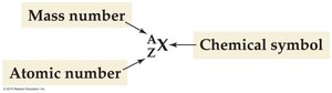
Isotopes are represented using a notation that includes the mass number, atomic number, and chemical symbol.
Notation: where A = mass number, Z = atomic number, X = chemical symbol.
Protons, neutrons, and electrons can be represented similarly.
Alpha Radiation
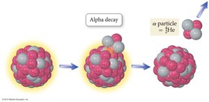
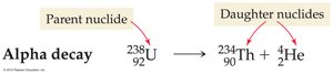
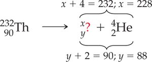
Alpha radiation occurs when an unstable nucleus emits an alpha particle, which consists of two protons and two neutrons.
Alpha particle symbol:
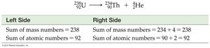
Example: Uranium-238 undergoes alpha decay to form thorium-234.
Alpha particles have high ionizing power but low penetrating power.
Beta Radiation
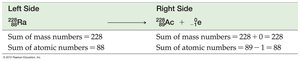
Beta radiation involves the emission of an electron from the nucleus as a neutron transforms into a proton. The atomic number increases by one.
Beta particle symbol:
Example: Radium-228 undergoes beta decay to form actinium-228.
Beta particles have moderate ionizing and penetrating power.

Gamma Radiation
Gamma radiation is electromagnetic radiation emitted from the nucleus, often accompanying other types of decay. Gamma rays have no mass or charge.
Gamma ray symbol:
Gamma rays have low ionizing power but high penetrating power.

Positron Emission
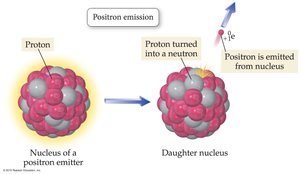
Positron emission occurs when a proton in the nucleus turns into a neutron, emitting a positron. The atomic number decreases by one.
Positron symbol:
Example: Phosphorus-30 undergoes positron emission to form silicon-30.
Positron emission has similar ionizing and penetrating power to beta emission.

Summary Table: Types of Radioactive Decay
The following table summarizes the main types of radioactive decay, their ionizing and penetrating power, and examples.
Decay Mode | Process | Ionizing Power | Penetrating Power | Example |
|---|---|---|---|---|
Alpha (α) | Emission of 2 protons and 2 neutrons | High | Low | |
Beta (β) | Neutron becomes a proton, electron emitted | Moderate | Moderate | |
Gamma (γ) | Photon emission | Low | High | |
Positron | Proton becomes a neutron, positron emitted | Moderate | Moderate |
Detecting Radioactivity
Film-Badge Dosimeters
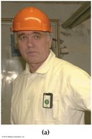
Film-badge dosimeters are used to monitor exposure to radiation by measuring the degree of exposure of photographic film.
Worn by personnel working with radioactive materials.
Degree of film exposure correlates with radiation dose.
Geiger-Müller Counter
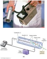
The Geiger-Müller counter detects energetic particles by ionizing argon gas in a chamber, producing an electrical signal.
Used for instantaneous detection of radioactivity.
Produces audible clicks corresponding to detected particles.
Natural Radioactivity and Half-Life
Background Radiation
Radioactive elements are naturally present in the environment due to their long half-lives or continuous formation. All nuclei with atomic numbers greater than 83 are unstable and radioactive.
Sources include ground, food, and cosmic rays.
Humans have adapted to background radiation.
The Concept of Half-Life
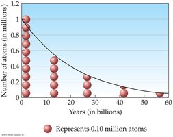
The half-life is the time required for half of the parent nuclides in a sample to decay. This concept is crucial for understanding radioactive decay rates.
After one half-life, 50% of the original sample remains.
After two half-lives, 25% remains; after three, 12.5% remains.
Decay Rates of Different Nuclides
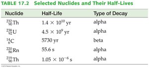
Different nuclides have unique half-lives, unaffected by physical or chemical conditions.
Thorium-232: 14 billion years
Radon-220: 1 minute


Table: Selected Nuclides and Their Half-Lives
Nuclide | Half-Life | Type of Decay |
|---|---|---|
yr | alpha | |
yr | alpha | |
5730 yr | beta | |
55.6 s | alpha | |
s | alpha |
Radioactive Decay Series
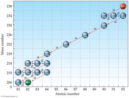
Uranium-238 undergoes a series of decays, eventually forming stable lead-206. Each step involves different types of radioactive decay and nuclides with varying half-lives.
Decay series includes alpha and beta emissions.
Radon gas is a product in the series, posing health risks.
Environmental Radon
Radon is a radioactive gas produced from uranium decay. It can accumulate indoors, increasing lung cancer risk.
Radon-222 has a half-life of 3.8 days.
High radon levels require ventilation or mitigation systems.

Applications of Radioactivity
Radiocarbon Dating
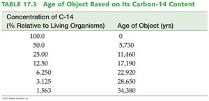
Radiocarbon dating uses the decay of carbon-14 to estimate the age of fossils and artifacts. Living organisms maintain a constant level of carbon-14, which decays after death.
Carbon-14 is formed by neutron bombardment of nitrogen in the atmosphere.
Half-life of carbon-14: 5730 years.
Age estimation is based on the remaining concentration of carbon-14.
Nuclear Fission and Fusion
Nuclear Fission
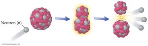
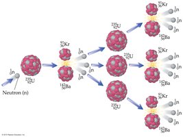
Nuclear fission is the splitting of a heavy nucleus into smaller fragments, releasing energy. Fission of U-235 produces neutrons that can initiate further fission, leading to a chain reaction.
Fission is used in nuclear power plants and atomic bombs.
Chain reactions require a critical mass of fissile material.
Nuclear Power Generation
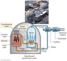
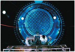
Nuclear power plants use fission to generate heat, which produces steam to drive turbines and generate electricity. Control rods regulate the chain reaction by absorbing neutrons.
Enriched U-235 fuel rods are used.
Control rods can be inserted or retracted to control the reaction.
Nuclear Accidents
Accidents at nuclear power plants, such as Chernobyl and Fukushima, highlight the risks of overheating and radiation release. Safety features are designed to prevent such incidents.
Chernobyl: Graphite core fire, widespread radioactive contamination.
Fukushima: Tsunami-induced cooling failure, partial meltdown.
US plants have containment structures for safety.
Nuclear Fusion
Nuclear fusion combines two light nuclei to form a heavier one, releasing energy. Fusion powers stars and hydrogen bombs, but requires extremely high temperatures.
Fusion reaction:
Fusion is not yet viable for electricity generation due to material limitations.
Effects of Radiation on Life
Acute Radiation Damage
High doses of radiation in a short period can kill cells, especially rapidly dividing ones, leading to weakened immune systems and potentially death.
Sources: nuclear bombs, exposed reactor cores.
Recovery possible in milder cases; severe cases can be fatal.
Increased Cancer Risk
Long-term exposure to lower doses of radiation can damage DNA, increasing cancer risk. Damaged DNA may cause abnormal cell growth and tumor formation.
Cancer risk increases with radiation exposure.
Thresholds for risk are difficult to determine.
Genetic Defects
Radiation can cause genetic defects in offspring if reproductive cells are affected. Such defects have been observed in laboratory animals but not conclusively in humans.
Measuring Radiation Exposure
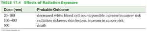
Radiation exposure is measured in units such as the curie, roentgen, and rem. The rem accounts for the ionizing power of different radiation types.
Average annual exposure: about one-third of a rem.
Radon is the largest source of natural exposure.
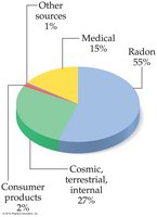
Table: Effects of Radiation Exposure
Dose (rem) | Probable Outcome |
|---|---|
20–100 | Decreased white blood cell count; possible increase in cancer risk |
100–400 | Radiation sickness; skin lesions; increase in cancer risk |
500 | Death |
Radioactivity in Medicine
Isotope Scans and Radiotherapy
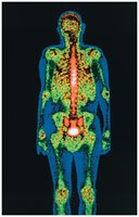
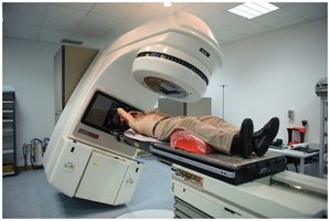
Radioisotopes are used in medical imaging and cancer treatment. Technetium-99, phosphorus-32, and iodine-131 are commonly used for diagnostic scans. Radiotherapy exposes tumors to gamma rays to destroy cancer cells.
Isotope scans detect bone and thyroid disorders.
Radiotherapy targets tumors while minimizing damage to healthy tissue.
Chapter Review and Learning Objectives
Understand the nature and discovery of radioactivity.
Write nuclear equations for alpha, beta, and positron decay.
Use the concept of half-life to solve problems.
Apply radiocarbon dating to estimate the age of artifacts.
Describe the effects of radiation on living organisms.
Explain the principles of nuclear fission and fusion.
Discuss the applications of radioactivity in medicine and power generation.
