 Back
BackDigestion and Absorption in the Small and Large Intestine
Study Guide - Smart Notes
 Tailored notes based on your materials, expanded with key definitions, examples, and context.
The Small Intestine
Gross Anatomy
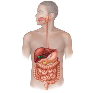
The small intestine is the principal organ for digestion and absorption of nutrients. It extends from the pyloric sphincter to the ileocecal valve, measuring approximately 2–4 meters in length and 2.5–4 cm in diameter. It is subdivided into three regions: the duodenum, jejunum, and ileum.
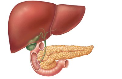
Duodenum: The first section (~25 cm), curves around the pancreas and receives digestive secretions from the liver and pancreas.
Jejunum: The middle section (~2.5 m), primarily involved in absorption.
Ileum: The final section (~3.6 m), joins the large intestine at the ileocecal valve.
Blood and Nerve Supply
Blood supply: Provided by the superior mesenteric artery; nutrient-rich blood is drained via the hepatic portal vein to the liver.
Nerve supply: Parasympathetic innervation via the vagus nerve and sympathetic innervation from thoracic splanchnic nerves.
Microscopic Anatomy
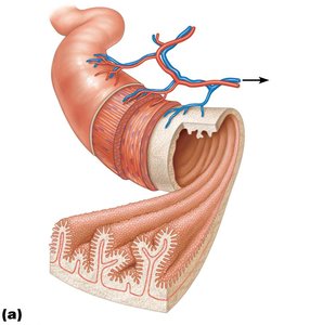
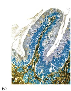
The small intestine is highly specialized for absorption, with structural modifications that increase its surface area approximately 600-fold to about 200 m2 (the size of a tennis court).
Circular folds: Permanent folds (~1 cm deep) that force chyme to spiral, slowing its movement and increasing absorption time.
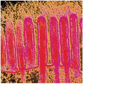
Villi: Fingerlike projections (~1 mm high) containing capillaries and a lymphatic vessel (lacteal) for nutrient absorption.
Microvilli: Cytoplasmic extensions forming the "brush border," which contains enzymes for final digestion of carbohydrates and proteins.
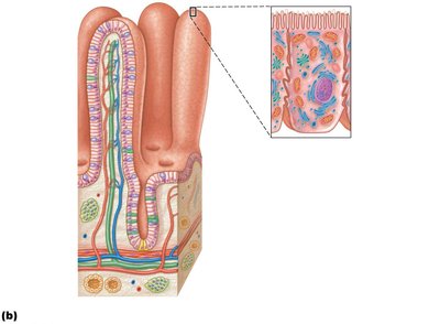
Cell Types in the Small Intestine
Enterocytes: Absorptive cells with microvilli; absorb nutrients and electrolytes.
Goblet cells: Secrete mucus for lubrication and protection.
Enteroendocrine cells: Release hormones (e.g., CCK, secretin) that regulate digestion.
Paneth cells: Secrete antimicrobial agents (defensins, lysozyme) to protect against bacteria.
Stem cells: Continuously divide to replenish the epithelium (renewed every 2–4 days).
Intestinal Juice
1–2 L secreted daily in response to distension or irritation.
Major stimulus: hypertonic or acidic chyme.
Slightly alkaline, isotonic with blood plasma, and mainly composed of water and mucus.
Digestive Processes in the Small Intestine
Chyme from the stomach contains partially digested carbohydrates and proteins, and undigested fats. The small intestine completes digestion and absorbs most nutrients and water within 3–6 hours.
Enzymes: Bile, bicarbonate, and digestive enzymes from the liver and pancreas aid digestion; brush border enzymes complete the process.
Regulation: Chyme entry is regulated to prevent osmotic imbalance and to ensure proper mixing with bile and pancreatic juice.
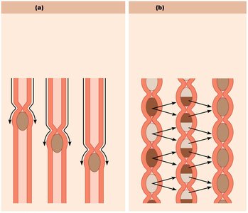
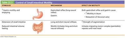
Motility of the Small Intestine
Segmentation: The most common motion after a meal, mixing and moving contents toward the ileocecal valve.
Peristalsis: Occurs between meals, moving remnants toward the large intestine (migrating motor complex).
Ileocecal valve: Controls entry of chyme into the large intestine, regulated by reflexes and hormones.
Stimulus | Mechanism | Effect on Motility |
|---|---|---|
Gastric motility and emptying | Gastroileal reflex (long neural reflex); Gastrin | Increased motility in ileum; relaxation of ileocecal valve |
Distension of small intestine | Long and short neural reflexes | Increased strength of segmentation |
Reduced intestinal volume; fasting | Motilin (initiates long and short neural reflexes) | Initiates migrating motor complex (peristaltic waves) until next meal |
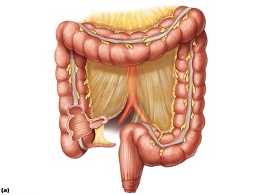
The Large Intestine
Gross Anatomy
The large intestine absorbs water and electrolytes, forms feces, and eliminates waste. It has unique features such as teniae coli (longitudinal muscle bands), haustra (pouches), and epiploic appendages (fat-filled pouches).
Cecum: First part, receives chyme from the ileum.
Appendix: Contains lymphoid tissue; may help recolonize gut bacteria.
Colon: Ascending, transverse, descending, and sigmoid regions.
Rectum: Contains valves to separate feces from gas.
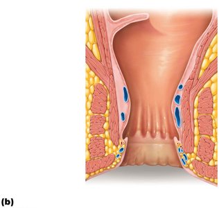
Anal canal: Opens to the exterior; has internal (smooth muscle) and external (skeletal muscle) sphincters.
Microscopic Anatomy
Thicker mucosa with simple columnar epithelium (except anal canal, which is stratified squamous).
No circular folds, villi, or digestive secretions; abundant goblet cells produce mucus.
Anal columns and sinuses aid in emptying and demarcate sensory regions (pectinate line).
Bacterial Flora
Over 1000 species of bacteria, outnumbering human cells 10:1.
Functions: Ferment indigestible carbohydrates, synthesize B vitamins and vitamin K, suppress pathogenic bacteria, and interact with the immune system.
Gut bacteria influence body weight, disease susceptibility, and even mood.
Digestive Processes in the Large Intestine
Residue remains for 12–24 hours; no further food breakdown except by bacteria.
Absorbs vitamins, water, and electrolytes; main function is propulsion and defecation.
Motility includes haustral contractions (slow, segmenting) and mass movements (powerful peristaltic waves).
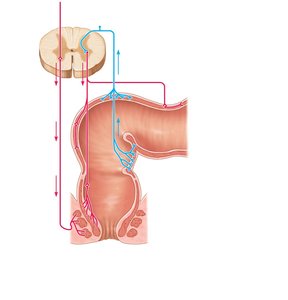
Defecation is triggered by rectal distension, involving both involuntary and voluntary muscle control.
Mechanisms of Digestion and Absorption
Enzymatic Hydrolysis
Digestion is a catabolic process where enzymes break down macromolecules into absorbable monomers by adding water (hydrolysis).
Absorption Mechanisms
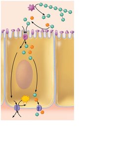
Absorption moves nutrients from the gut lumen into the body, primarily through enterocytes.
Lipid molecules diffuse passively; other nutrients require active transport.
Most absorption occurs before chyme reaches the ileum.
Digestion and Absorption of Nutrients
Carbohydrates
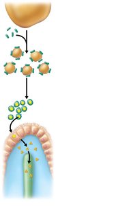
Only monosaccharides (glucose, fructose, galactose) are absorbed.
Starch digestion begins with salivary amylase, continues with pancreatic amylase, and is completed by brush border enzymes.
Monosaccharides are absorbed via cotransport with Na+ (secondary active transport) and facilitated diffusion.

Proteins
Digestion begins in the stomach (pepsin), continues in the small intestine (pancreatic proteases), and is completed by brush border enzymes.
Amino acids are absorbed via cotransport with Na+ or H+ and facilitated diffusion.
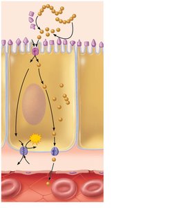
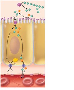
Lipids
Emulsified by bile salts, digested by pancreatic lipases into monoglycerides and fatty acids.
Products form micelles, diffuse into enterocytes, are reassembled into triglycerides, and packaged as chylomicrons for lymphatic transport.
Short-chain fatty acids diffuse directly into blood.
Nucleic Acids
Digested by pancreatic nucleases and brush border enzymes into nitrogenous bases, pentose sugars, and phosphate ions.
Absorbed by active transport into blood.
Absorption of Vitamins, Electrolytes, and Water
Vitamins: Fat-soluble (A, D, E, K) absorbed with micelles; water-soluble (C, B) by diffusion or transporters; B12 requires intrinsic factor.
Electrolytes: Most actively absorbed; iron and calcium absorption regulated by need.
Water: Absorbed by osmosis, mainly in the small intestine (95%).
Clinical Considerations
Lactose intolerance: Deficiency of lactase leads to osmotic diarrhea and gas production.
Malabsorption syndromes: Can result from impaired bile or pancreatic juice delivery, or damaged mucosa (e.g., celiac disease).
Appendicitis: Inflammation of the appendix, potentially leading to peritonitis if ruptured.
Antibiotic-associated diarrhea: Overgrowth of Clostridium difficile after antibiotic use can cause severe colitis.
Diverticulosis/Diverticulitis: Outpouchings of the colon wall, which can become inflamed and rupture.
Irritable bowel syndrome: Functional disorder with abdominal pain and altered bowel habits, often stress-related.
