Indietro
A&P II Exam 1 Study Guide: Blood, Cardiovascular, Respiratory, and Immune Systems
Guida di studio - Note intelligenti
 Appunti personalizzati basati sui tuoi materiali, ampliati con definizioni chiave, esempi e contesto.
Blood Anatomy and Physiology
Blood Components and Functions
The blood is a specialized connective tissue composed of cells suspended in plasma. It plays a critical role in transport, regulation, and protection within the body.
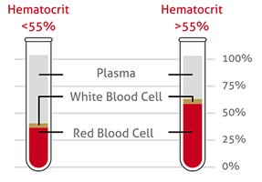
Hematocrit: The percentage of packed red cell volume in a whole blood sample. Normal values: males 42-52%, females 37-47%.
Erythrocyte: Red blood cell responsible for oxygen transport and determining blood type.
Hemoglobin: Oxygen-carrying pigment in erythrocytes. Normal levels: males 13.5-18 g/100ml, females 12-16 g/100ml.
Buffy coat: Layer of white blood cells between plasma and red blood cells; involved in immune functions.
Plasma: Liquid component of blood; distributes materials, regulates pH, and contains antibodies.
Anemia: Lower than normal hematocrit.
Polycythemia: Higher than normal hematocrit, often seen at high altitudes.
Example: A drop in red blood cell number triggers the kidney to release erythropoietin, increasing red blood cell production in bone marrow.
Blood Typing and Genetics
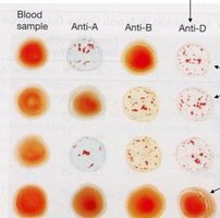
Blood type is determined by the presence of glycoprotein antigens on erythrocyte surfaces. The ABO system is inherited, with three genes (A, B, O) coding for blood types.
Type A: Antigen A present, B absent.
Type B: Antigen B present, A absent.
Type AB: Both antigens present.
Type O: Both antigens absent.
Rh group: The "D" antigen is most important. Rh+ individuals have the D antigen; Rh- do not. Rh antibodies are formed after exposure, which can cause hemolytic anemia in newborns if the mother is Rh- and the baby is Rh+.
Blood Type | Antigen A | Antigen B |
|---|---|---|
A | Present | Absent |
B | Absent | Present |
AB | Present | Present |
O | Absent | Absent |
Hemoglobin and Oxygen Transport
Hemoglobin is a protein with four heme groups, each containing iron that binds oxygen. It forms oxyhemoglobin when combined with oxygen.
Elevated hemoglobin: Polycythemia, congestive heart failure, chronic obstructive pulmonary disease, high altitudes.
Decreased hemoglobin: Anemia, hyperthyroidism.
Antigens, Agglutinins, and Transfusion Reactions
Antigens are glycoproteins on erythrocyte membranes. Agglutinins are antibodies in plasma that cause agglutination and lysis if incompatible blood is transfused.
Universal donor: O-
Universal recipient: AB+
Cardiovascular System
Heart Anatomy
The heart is a muscular organ with four chambers and valves that ensure one-way blood flow. Review the Heart Atlas for detailed structure.
Cardiac Physiology
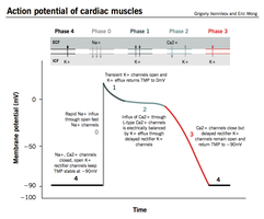
Cardiac muscle cells exhibit autorhythmicity, allowing them to generate action potentials without external stimuli. The refractory period prevents tetanus and wave summation.
Autorhythmicity: Cardiac cells generate action potentials independently.
Absolute refractory period: No action potential can be generated, lasting 200-250 ms.
Wave summation: Not possible in cardiac muscle due to long refractory period.
Cardiac Arrhythmias
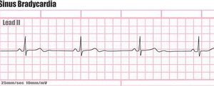
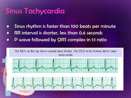
Abnormal heart rhythms include extrasystoles, ventricular fibrillation, bradycardia, and tachycardia.
Ventricular fibrillation: Disorganized electrical activity, ineffective pumping.
Bradycardia: Slow heart rate.
Tachycardia: Fast heart rate.
Autonomic Regulation of Heart Rate
The sympathetic nervous system increases heart rate and contractility, while the parasympathetic system decreases heart rate via the vagus nerve.
Sinoatrial node: Pacemaker of the heart.
Vagus nerve: Excessive stimulation can stop the heart; cutting it results in autonomous SA node rhythm.
Blood Vessel Physiology
Blood vessel radius is controlled by smooth muscle contraction (vasoconstriction) and relaxation (vasodilation), affecting blood flow.
Laminar flow: Free-flowing blood in the vessel center.
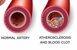
Atherosclerosis: Plaques increase resistance and decrease flow.
Arteriosclerosis: Loss of arterial elasticity.
Respiratory System
Respiratory Anatomy
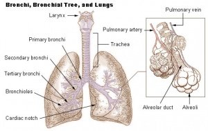
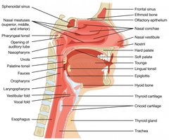
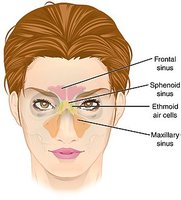
The respiratory system includes the nasal cavity, pharynx, larynx, trachea, bronchi, and lungs. The lungs contain multiple lobes and alveoli for gas exchange.
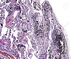
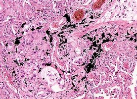
Histology of Lung Tissue
Normal lung tissue is characterized by thin alveolar walls and clear air spaces. Pathological changes include coal dust accumulation and cancer.
Respiratory Physiology
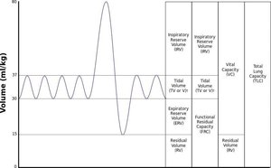
Ventilation involves inspiration and expiration. Lung volumes and capacities are measured to assess pulmonary function.
Tidal volume (TV): Normal quiet breathing (500 ml).
Inspiratory reserve volume (IRV): Maximum air inspired (males 3100 ml, females 1900 ml).
Expiratory reserve volume (ERV): Maximum air expired (males 1200 ml, females 700 ml).
Residual volume (RV): Air remaining after expiration (males 1200 ml, females 1100 ml).
Forced vital capacity (FVC): Maximum air expired after maximum inspiration.
Gas Exchange in Alveoli
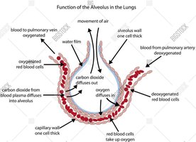
Gas exchange occurs in alveoli, where oxygen diffuses into blood and carbon dioxide diffuses out. The alveolar wall is one cell thick, facilitating efficient exchange.
Respiratory Diseases
Emphysema: Loss of elastic recoil, difficulty expiring.
Asthma: Bronchiole smooth muscle spasm, airway constriction.
Pneumothorax: Equal intrapleural and atmospheric pressure, lung collapse.
Immune System
Lymphocyte Development and Activation
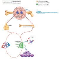
Lymphocytes originate in red bone marrow and mature into B and T cells. Activation occurs upon exposure to antigens.
Antibody Structure
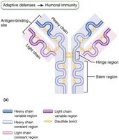
Antibodies are proteins with variable and constant regions, composed of light and heavy chains. They bind antigens and mediate immune responses.
ELISA (Enzyme Linked Immunosorbent Assay)
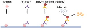
ELISA is a laboratory technique used to detect the presence of antigens or antibodies in a sample.
Add sample to tube; proteins bind to the tube.
Add primary antibody; binds to antigen of interest.
Add secondary antibody labeled with enzyme; binds to primary antibody.
Add substrate; color change indicates presence of antigen.
Lab Safety
GHS Pictograms
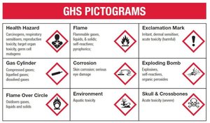
The Globally Harmonized System (GHS) pictograms are used to identify hazards in the laboratory, including health, flammability, and environmental risks.