 Indietro
Anatomy & Physiology Study Guide: Endocrine System, Blood, and Cardiovascular System
Guida di studio - Note intelligenti
 Appunti personalizzati basati sui tuoi materiali, ampliati con definizioni chiave, esempi e contesto.
Chapter 13 – Endocrine System
General Characteristics of the Endocrine System
The endocrine system consists of glands that secrete hormones directly into the bloodstream, regulating various physiological processes. Understanding the differences between exocrine and endocrine glands, hormone action, and hormone regulation is fundamental to grasping endocrine physiology.
Exocrine vs. Endocrine Glands:
Exocrine glands release their products (e.g., enzymes, sweat) into ducts that lead to target sites.
Endocrine glands secrete hormones directly into the bloodstream, affecting distant target cells.
Hormone: A chemical messenger released by endocrine glands that alters the activity of specific target cells.
Target Cell: A cell with specific receptors for a hormone, enabling it to respond to that hormone.
Regulation of Sensitivity:
Up-regulation: Increase in receptor number, enhancing sensitivity to a hormone.
Down-regulation: Decrease in receptor number, reducing sensitivity.
Chemical Classes of Hormones:
Steroid hormones: Derived from cholesterol; examples include cortisol and estrogen.
Amine hormones: Derived from amino acids; examples include epinephrine and thyroxine.
Peptide/protein hormones: Chains of amino acids; examples include insulin and growth hormone.
Hydrophobic Hormones: Steroid and thyroid hormones; receptors are located inside the cell (cytoplasm or nucleus). They alter gene expression.
Hydrophilic Hormones: Peptide/protein and most amine hormones; receptors are on the cell membrane. They activate second messenger systems.
Lipophilic: Fat-soluble (hydrophobic); Lipophobic: Water-soluble (hydrophilic).
Mechanisms for Controlling Hormone Release:
Humoral stimuli (changes in blood levels of ions/nutrients)
Neural stimuli (nerve impulses)
Hormonal stimuli (other hormones)
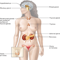
Hypothalamus and Pituitary Gland
The hypothalamus and pituitary gland are central regulators of endocrine function, controlling the release of many hormones that affect growth, metabolism, and homeostasis.
Pituitary Gland Location: Base of the brain, connected to the hypothalamus by the infundibulum.
Histology:
Anterior pituitary: Glandular tissue.
Posterior pituitary: Neural tissue.
Hormones Released:
Anterior pituitary: Growth hormone (GH), thyroid-stimulating hormone (TSH), adrenocorticotropic hormone (ACTH), follicle-stimulating hormone (FSH), luteinizing hormone (LH), prolactin (PRL).
Posterior pituitary: Antidiuretic hormone (ADH), oxytocin.
Portal System: Network of blood vessels connecting hypothalamus to anterior pituitary, allowing hormone transport.
Hormonal Imbalances:
Pituitary dwarfism: GH deficiency in childhood.
Pituitary giantism: GH excess in childhood.
Acromegaly: GH excess in adulthood.
Diabetes insipidus: ADH deficiency, causing excessive urination.
Thyroid and Parathyroid Glands
The thyroid and parathyroid glands regulate metabolism and calcium homeostasis through hormone secretion.
Thyroid Follicles: Release thyroxine (T4) and triiodothyronine (T3), which increase metabolic rate.
Hyperthyroidism Symptoms: Weight loss, increased heart rate, anxiety.
Hypothyroidism Symptoms: Weight gain, fatigue, cold intolerance.
Graves Disease: Autoimmune hyperthyroidism.
Simple Goiter: Thyroid enlargement due to iodine deficiency.
Extrafollicular Cells: Release calcitonin, lowering blood calcium.
Parathyroid Glands: Release parathyroid hormone (PTH), increasing blood calcium.
Adrenal Glands
The adrenal glands produce hormones that regulate stress response, metabolism, and electrolyte balance.
Adrenal Medulla: Releases epinephrine and norepinephrine (fight-or-flight response).
Adrenal Cortex: Releases cortisol (metabolism), aldosterone (electrolyte balance), and androgens.
Cushing’s Syndrome: Excess cortisol, causing obesity, hypertension, and muscle weakness.
Pancreas, Pineal Gland, and Thymus
These glands regulate blood glucose, circadian rhythms, and immune function.
Pancreas: Both exocrine (digestive enzymes) and endocrine (hormones) functions.
Islets of Langerhans: Groups of cells releasing hormones.
Alpha Cells: Release glucagon (raises blood glucose).
Beta Cells: Release insulin (lowers blood glucose).
Diabetes Mellitus:
Type 1: Autoimmune destruction of beta cells.
Type 2: Insulin resistance.
Symptoms: Polyuria, polydipsia, polyphagia.
Pineal Gland: Releases melatonin (regulates sleep-wake cycle).
Thymus: Releases thymosins (stimulate T-cell development).
Chapter 14 – Blood
Characteristics of Blood
Blood is a specialized connective tissue composed of plasma and formed elements, playing a vital role in transport, regulation, and protection.
Blood Plasma: Liquid component; contains water, proteins, nutrients, hormones, and waste.
Formed Elements: Erythrocytes (RBCs), leukocytes (WBCs), platelets.
Hematocrit: Percentage of RBCs in blood.
Buffy Coat: Layer containing WBCs and platelets after centrifugation.
Plasma Proteins:
Albumins: Maintain osmotic pressure; made in liver.
Globulins: Transport and immune functions; made in liver and lymphoid tissue.
Fibrinogen: Clotting; made in liver.
Red Blood Cells
Red blood cells (erythrocytes) are responsible for oxygen and carbon dioxide transport. Their production and destruction are tightly regulated.
Hematopoiesis: Formation of blood cells; occurs in yolk sac, liver, spleen (before birth), and red bone marrow (after birth).
Hemocytoblast: Stem cell giving rise to all blood cells.
Myeloid Line: Produces RBCs, platelets, granulocytes, monocytes.
Lymphoid Line: Produces lymphocytes.
Erythrocyte Characteristics: Biconcave, anucleate, flexible.
Hemoglobin: Four globin chains, each with a heme group; binds oxygen and carbon dioxide.
Each hemoglobin molecule can transport up to four oxygen molecules.
Reticulocytes: Immature RBCs.
Normal RBC Count: 4.5–6 million/μL.
Erythropoietin: Hormone from kidneys and liver stimulating RBC production.
RBC Lifespan: ~120 days.
Dietary Factors: Iron, vitamin B12, folic acid.
Anemia Types: Iron-deficiency, pernicious, sickle cell, aplastic, hemolytic.
Sickle Cell Disease: Genetic mutation causing abnormal hemoglobin and RBC shape.
RBC Removal: Spleen and liver; hemoglobin broken down into heme (bilirubin) and globin (amino acids).
Jaundice: Accumulation of bilirubin.
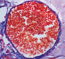
White Blood Cells
White blood cells (leukocytes) are essential for immune defense and are classified as granulocytes or agranulocytes.
Granulocytes: Neutrophils, eosinophils, basophils.
Agranulocytes: Lymphocytes, monocytes.
Normal WBC Count: 4,000–11,000/μL.
Differential WBC Count: Percentage of each type of WBC.
Leukemia: Cancer of WBCs; acute or chronic types.
Platelets and Hemostasis
Platelets (thrombocytes) are cell fragments involved in blood clotting. Hemostasis is the process of stopping bleeding.
Platelet Structure: Small, anucleate cell fragments.
Origin: Megakaryocytes in bone marrow.
Normal Platelet Count: 150,000–400,000/μL.
Phases of Hemostasis:
Vascular spasm
Platelet plug formation
Coagulation (clotting)
Intrinsic vs. Extrinsic Pathways: Both lead to conversion of fibrinogen to fibrin.
Substances Required: Calcium, clotting factors, vitamin K.
Positive Feedback: Clot formation amplifies itself.
Clot Dissolution: Plasmin enzyme.
Clotting Disorders: Hemophilia (factor deficiency), thrombus (stationary clot), embolus (traveling clot), embolism (blocked vessel).
Blood Types and Blood Transfusions
Blood types are determined by antigens and antibodies, affecting transfusion compatibility.
ABO Blood Group:
A: A antigen, anti-B antibody
B: B antigen, anti-A antibody
AB: A and B antigens, no antibodies (universal recipient)
O: No antigens, anti-A and anti-B antibodies (universal donor)
Rh Factor: Rh+ (antigen present), Rh- (antigen absent).
Hemolytic Disease of the Newborn: Rh incompatibility between mother and fetus.
Chapter 15 – Cardiovascular System
General Characteristics, Heart Anatomy, and Blood Flow
The cardiovascular system consists of the heart and blood vessels, responsible for circulating blood throughout the body. Understanding heart anatomy and blood flow is essential for grasping cardiovascular physiology.
Pulmonary Circuit: Carries blood from heart to lungs and back.
Systemic Circuit: Carries blood from heart to body tissues and back.
Pericardium Layers: Fibrous, parietal, visceral (epicardium).
Heart Layers: Epicardium, myocardium, endocardium.
Right Side of Heart: Deoxygenated blood.
Left Side of Heart: Oxygenated blood.
Blood Vessels to Right Atrium: Superior vena cava, inferior vena cava, coronary sinus.
Blood Flow Path: Right atrium → right ventricle → pulmonary artery → lungs → pulmonary vein → left atrium → left ventricle → aorta.
Fetal Circulation Remnants: Foramen ovale, ductus arteriosus.
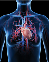
Coronary Circulation
Coronary arteries and veins supply the heart muscle with oxygen and nutrients.
Angina Pectoris: Chest pain due to reduced blood flow.
Myocardial Infarction: Heart attack; death of heart tissue.
Cardiac Conduction System and ECGs
The cardiac conduction system coordinates heart contractions, and ECGs record electrical activity.
Systole: Contraction phase.
Diastole: Relaxation phase.
Cardiac Cycle: Complete heartbeat.
Cardiac Muscle Cells: Intercalated discs allow functional syncytium.
Conduction System: SA node, AV node, bundle of His, bundle branches, Purkinje fibers.
Normal Pacemaker: SA node.
ECG Deflections: P wave (atrial depolarization), QRS complex (ventricular depolarization), T wave (ventricular repolarization).
Arrhythmias: Fibrillation, tachycardia, bradycardia, flutter, ectopic pacemaker.
The Cardiac Cycle
The cardiac cycle involves changes in chamber volume and pressure, determining blood flow and valve operation.
Pressure-Volume Relationship: Increased volume decreases pressure; decreased volume increases pressure.
Valve Operation: Valves open/close based on pressure differences.
Heart Sounds: S1 (lubb) – AV valves close; S2 (dupp) – semilunar valves close.
Heart Murmur: Abnormal sound due to valve issues.
Sympathetic/Parasympathetic Effects: Sympathetic increases heart rate; parasympathetic decreases heart rate.
Parasympathetic Nerve: Vagus nerve.
Baroreceptors: Located in carotid sinus and aortic arch; detect blood pressure changes.
Cardiac Center Location: Medulla oblongata.
Body Temperature: Increased temperature raises heart rate.
Blood Vessels
Blood vessels transport blood throughout the body, with arteries, veins, and capillaries having distinct structural and functional characteristics.
Sequence in Systemic Circuit: Arteries → arterioles → capillaries → venules → veins.
Blood Vessel Layers: Tunica intima, tunica media, tunica externa.
Arteries: Thick walls, high pressure.
Capillaries: Single layer of endothelial cells.
Exchange Methods: Diffusion, filtration, osmosis, transcytosis.
Filtration/Reabsorption: Driven by hydrostatic and osmotic pressures.
Disruptions: Heart failure, liver failure, immune reactions.
Veins: Thin walls, valves, low pressure.
Blood Movement in Veins: Skeletal muscle contraction, respiratory movements, valves.
Vascular Disorders: Atherosclerosis, arteriosclerosis, aneurysm, varicose veins.
Blood Pressure
Blood pressure is the force exerted by blood on vessel walls, influenced by cardiac output, blood volume, and peripheral resistance.
Blood Pressure: Usually refers to arterial pressure.
Blood Pressure Reading: Systolic/diastolic; pulse pressure = systolic - diastolic; mean arterial pressure = diastolic + 1/3 pulse pressure.
Cardiac Output: Volume of blood pumped per minute. $CO = HR \times SV$
Stroke Volume: Volume ejected per beat.
Frank-Starling Law: Increased venous return increases stroke volume.
Blood Volume: Directly proportional to blood pressure.
Hormones Influencing Blood Volume: ADH, aldosterone, atrial natriuretic peptide.
Peripheral Resistance: Resistance to blood flow; affected by vessel diameter, blood viscosity, and vessel length.
Vessel Radius: Smaller radius increases resistance and blood pressure.
Sympathetic Effects: Vasoconstriction increases blood pressure.
Hormonal Effects:
Epinephrine: Vasoconstriction
Angiotensin II: Vasoconstriction
Atrial Natriuretic Peptide: Vasodilation
Histamine: Vasodilation
Blood Pressure Regulation: Baroreceptor reflex, hormonal control.
Hypertension: Chronic high blood pressure.
Systemic Arteries and Veins
Major systemic arteries and veins are essential for distributing blood throughout the body and are commonly identified in laboratory exams.
Systemic Arteries: Aorta, carotid, subclavian, femoral, etc.
Systemic Veins: Superior/inferior vena cava, jugular, femoral, etc.
